
The Common Vein Copyright 2007
Introduction
At the histological level the pancreas is made up of compound glands in “bunch of grapes” fashion.
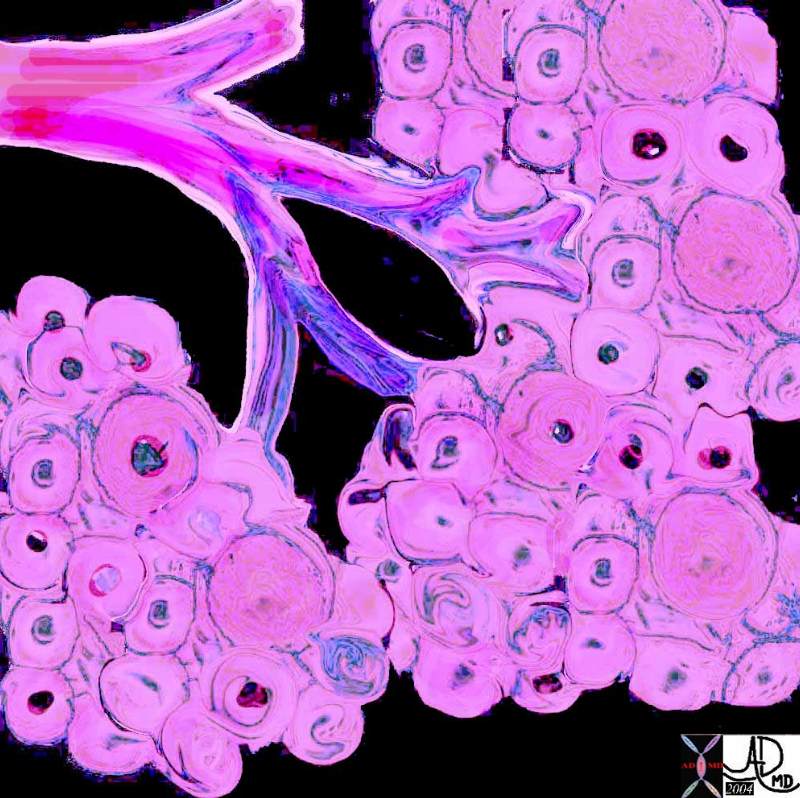 The pancreas has an exocrine and endocrine component. The exocrine compnent is demonstrated above in 3D with acini in “cluster of grapes” formation subtended by a duct. Courtesy Ashley Davidoff MD 32645a06
Conceptually the organization of functional units around a ductal system is the same as the lung but the “alveoli” are filled with solid cellular tissue rather than air. Interspersed among the clusters of exocrine glands are the sparse accumulations of Langerhans glands that represent only 2% of the volume of the gland. Between the glands is an interstitial fluid that is gel like in the young and with advancing age is replaced by fat that creeps in from the retroperitoneum. The little protection afforded by the flimsy pancreatic “capsule” makes this infiltration easy. Similarly when disease occurs in the pancreas there is little to hold the disease within the confines of the pancreas so that exudate or malignancy extends outward early in the natural history of the disease process. This has very important implications.
There are 20-35 secondary ducts that arise in herring bone fashion off the main duct. The acini accumulate around the ductules and their branches to form lobules
80% of the parenchyma consists of lobules of pancreatic acini that are reponsible for the exocrine secretions. Approximately 18% of the gland is derived from ductal system and intersttium ,while the islets of Langerhans makeup only 2% of the gland. Richly innervated peripancreatic areolar tissue penetrates the parenchyma as thin septa , dividing it up into lobules. Each lobule is drained by a small branch of the pancreatic duct. dips in between lobules.
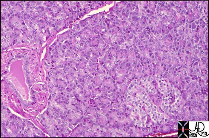
Normal histology of the pancreas showing the islet of Langerhans in the right lower corner of the image with the remaining glandular tissue being the exocrine portion of the gland. The islets only make up about 2% of the gland. A duct can be seen in the left lower quadrant surrounded by a thicklayer of connective tissue. Courtesy Barbara Banner 15282
Acini
The acini are rounded or have a short tubular form and consist of single rows of epithelial cells lying on a basal lamina. Lining the lumen of the acinus are pyramidal acinar cells. There are in addition unique cells called the centroacinar cells. The lumen of the acinus connects with the intralobular ducts to form the interlobular ducts. These ducts join to form the main pancreatic duct. Lining the ductules are columnar cells, goblet cells, and occasional argentaffin cells. The larger ducts are surrounded by a prominent layer of connective tissue.Thick layers of connective tissue and elastic fibers surround the large ducts.
Islets of Langerhans
The endocrine portion of the pancreas consists of about one million islets of Langerhans, which are distributed throughout the gland but are relatively concentrated in the tail of the pancreas. The islets are surrounded a rich capillary plexus.
About 75% – 80% of islet cells are insulin secreting beta cells, and they lie centrally in the islet. 10% -20% of the islet are alpha cells which lie in the periphery and secrete glucagon. Delta cells are interspersed throughout the islet, compose about 5% and secrete somatostatin.
At the histological level the pancreas is made up of compound glands in “bunch of grapes” fashion.
| The pancreas has an exocrine and endocrine component. The exocrine compnent is demonstrated above in 3D with acini in “cluster of grapes” formation subtended by a duct. Courtesy Ashley Davidoff MD 32645a06
Conceptually the organization of functional units around a ductal system is the same as the lung but the “alveoli” are filled with solid cellular tissue rather than air. Interspersed among the clusters of exocrine glands are the sparse accumulations of Langerhans glands that represent only 2% of the volume of the gland. Between the glands is an interstitial fluid that is gel like in the young and with advancing age is replaced by fat that creeps in from the retroperitoneum. The little protection afforded by the flimsy pancreatic “capsule” makes this infiltration easy. Similarly when disease occurs in the pancreas there is little to hold the disease within the confines of the pancreas so that exudate or malignancy extends outward early in the natural history of the disease process. This has very important implications. There are 20-35 secondary ducts that arise in herring bone fashion off the main duct. |