
The Common Vein Copyright 2007
Ashley Davidoff MD
Author Ashley Davidoff MD
Collaborators Charles Allison MD Adam Asarch MS II David Lee MD Scott Tsai MD
Overview
Serous cystadenoma (microcystic cystadenoma or glycogen-rich cystadenoma) is a benign and relatively uncommon condition of the pancreas (Pyke)(related articles) It is more common than its counterpart, mucinous cystadenoma by about 2 times. It is also about 2 times as common in women as in men. It most commonly presents in the 7th decade.
Imaging
The imaging features are by no means pathgnomonic but some features are characteristic. The average size is about 6cm but they range from a few cms. to as large as 20cms. They are most commonly situated in the head or neck of the pancreas. There is a central stellate scar that enhances late in the bolus. The central scar sometimes calcifies and forms a stellate or sunburst appearance. The matrix of the lesion is usually multicystic, with about 6 cysts, each usually measuring less than 2cms. It has been described as having a honeycomb appearance. The cysts are low density on the non contrast study but demonstrate septations with the contrast. MRI is useful in characterizing the hemorrhage that often occurs in the cysts. When the cysts are very small, US will show an echogenic mass due to the multiple interfaces created by the connective tissue walls that are closely packed. Through transmission though will reveal the innate cystic nature of the lesion.
Although the lesions are large at presentation they do not usually cause obstruction of the pancreatic duct or bile duct. (10-15%) The lack of communication with the pancreatic duct is unlike its mucinous counterpart, which often is in communication with the duct.
Approach
When a cystic lesion is identified in a patient, the first step is to exclude pancreatitis related disease – a pseudocyst, or a dilated pancreatic duct. When these entities have been excluded, the age, sex, location and morphology of the lesion are prime components for initial consideration. An elderly female with a cystic lesion in the body or neck, that has central stellate sunburst calcification with multicystic clusters of cysts, leads to serous cystadenoma being the prime suspect. If the lesion in addition has not changed in 2 years with an ERCP that demonstrates a normal sized duct with no communication to the duct, benign serous cystadenoma is even more likely. If the features are in question (which is a common occurrence), then biopsy and aspiration is required. The finding of a low viscosity fluid, with absence of mucin, and the presence of glycogen rich cells makes the diagnosis certain. In the absence of a definitive pathological diagnosis, it is prudent to follow these lesions because of the significant crossover with the findings of mucinous cystadenoma, and the potential of the latter entity to undergo malignant transformation.
Pathology
 This histological section of the pancreas is from the records of a patient with a serous cystadenoma. The lumen of the cyst is at the top of the image and is cream colored. A single layer of epithelial cells lines the cyst. A pseudocyst on the other hand would have no epithelial lining.15316b01Courtesy Barbara Banner MD
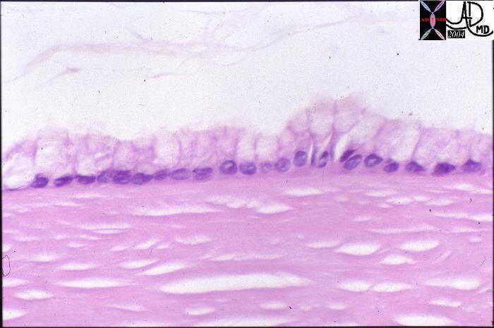 This high power view is of a histological section from the records of a patient with a serous cystadenoma of the pancreas. The lumen of the cyst is at the top of the image and is cream colored with floating debris. A single layer of columnar epithelial cells lines the cyst. A pseudocyst on the other hand would have no epithelial lining.15317 Courtesy Barbara Banner MD
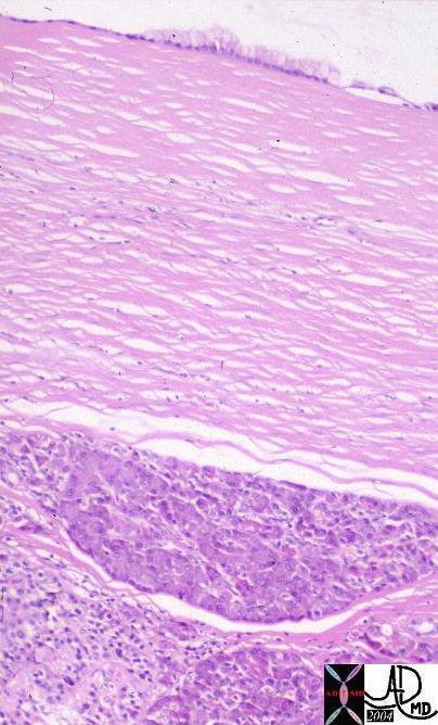
 This high powered histological section is from the records of a patient with a pseudocyst. At the top of the image the white space represents the cyst lumen. The lining is made of fibrous tissue rather than an epithelium. A true cyst is lined by epithelial cells. 15306 Courtesy Barbara Banner MD
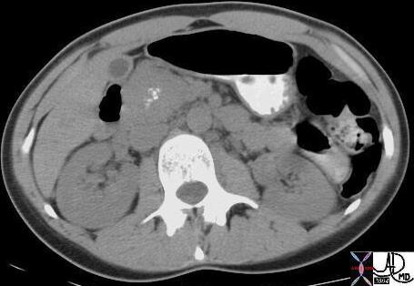
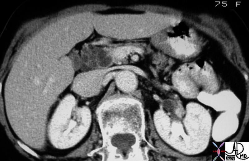 The CTscan through the head of the pancreas is of a 75 year old female with abdominal discomfort. There is a multicystic mass in the head of the pancreas with thickened walls and a Swiss cheese appearance to the lesion . The age sex, appearance, and location make a serous cystadenoma most likely. 40585 Courtesy Ashley Davidoff MD
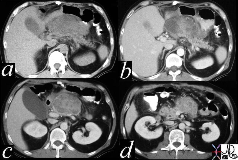
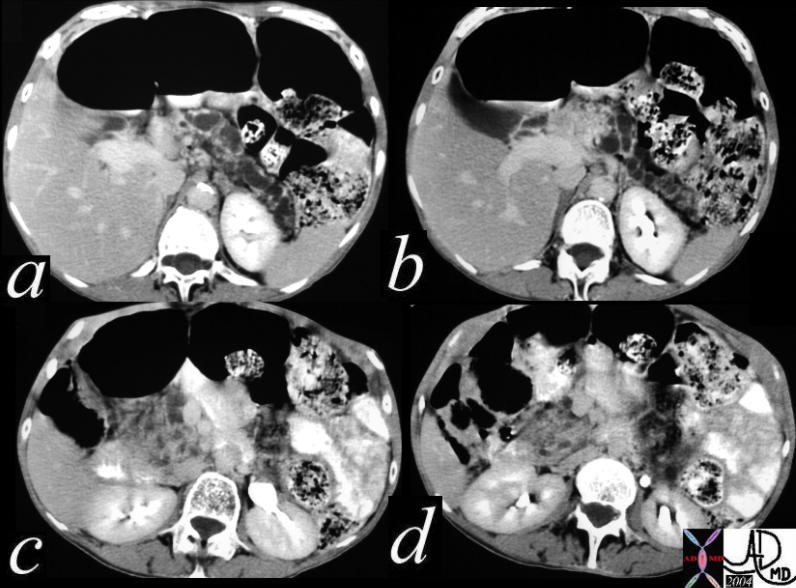
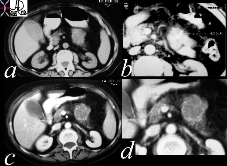 The series of CT scans through the abdomen are one years apart. The 78year old patient was first noted to have a low density enhancing mass (a,b) which was biopsied (b) and shown to have features that suggested a serous cystadenoma. One year later the multicystic mass was perhaps a little larger. At this time the multicystic features were better demonstrated. Although the pathology suggested a benign disease the slight apparent enlargement warrants continued follow up. 40536c Courtesy Ashley Davidoff MD
CTscan
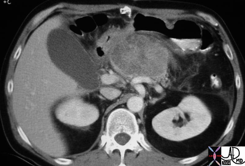 This CT scan of the abdomen shows a large enhancing mass in the body of the pancreas. The multicystic nature is not apparent. At surgery this was shown to be a serous cystadenoma. 40566 Courtesy Ashley Davidoff MD
 This CT scan of the abdomen shows series of a large enhancing mass in the body and neck of the pancreas. There is a large cyst in b, with smaller ill defined cystic lesions in c. At surgery and pathology this lesion was shown to be a serous cystadenoma. 40572c
Courtesy Ashley Davidoff MD
The enhancement pattern of the mass suggests a multicystic nature with faint septations seen in the next image.
 This abdominal CT scan without contrast is from the records of a patient with known von Hippel-Lindau syndrome. It shows a soft tissue density mass in the head of the pancreas with central stellate calcifications. While the central stellate type calcifications fit well with cystadenoma, the soft tissue nature does not. 18378 Courtesy Ashley Davidoff MD
 This abdominal CT scan with contrast is from the records of a patient with known von Hippel-Lindau syndrome. It shows a soft tissue density mass in the head of the pancreas that is relatively less dense than the soft tissues, with central calcifications and subtle compartmentalization by enhancing septations. Radiologically the lesion was felt to be more like a serous cystadenoma on this image. 19393 Courtesy Ashley Davidoff MD+ imaging radiology CTscan C-
 This series images is from the abdominal CT scan of an elderly woman with a stable multilcystic mass in the head of the pancreas. (c,d) The patient had been followed for several years without change. This history would fit a benign serous cystadenoma, but the pancreatic duct is, and has been dilated in the past(a,b) The latter feature is an uncommon finding occurring in 10-15% of patients with benign disease. Continued surveillance is therefore warranted. 40502c Courtesy Ashley Davidoff MD
Unusual Cases
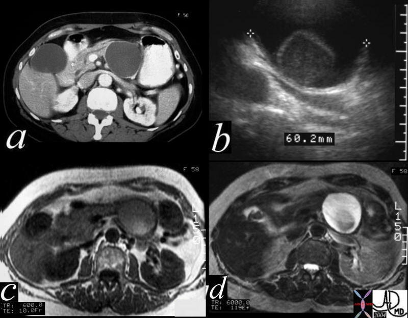 This CTscan is through the tail of the pancreas shows what appears to be a simple cyst (a), but which proves to have a mural nodule on ultrasound (b). On the T1 weighted image (c) the nodule shows high intensity suggesting increased protein or acute blood, and the T2 (d) reveals the cystic nature of the matrix. At pathology the lesion was a benign serous cystadenoma. The high intensity on T1 was due to the high protein in the mucin. 40487c01 Courtesy Ashley Davidoff MD
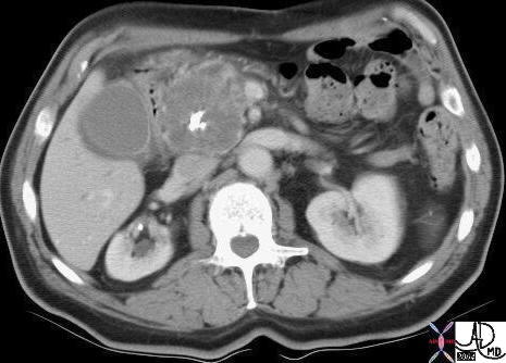
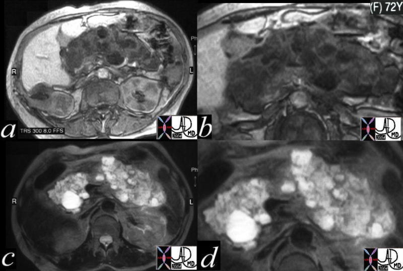 This CT scan in a 72 year old female through the pancreas shows a series of complex cystic and solid masses diffusely throughout the pancreas, with a few cystic masses in the right kidney. These findings are consistent with a diagnosis of von Hippel-Lindau syndrome in which the gamut of cystic and solid, benign and malignant tumors of the pancreas manifest. 24061c Courtesy Ashley Davidoff MD