
The Common Vein Copyright 2007
SHAPE OF THE PANCREAS
This section deals with the variety of shapes of the normal pancreas. The pancreas has been compared to many objects including an elongated comma on its side, an elongated number 9 on its side, a prism, a banana, an inverted and curved upside down tobacco pipe, and even an old fashioned revolver. Placing a seahorse or a woodpecker with head down and tail up probably brings us closest to the complex shape of the pancreas. The objects used for description are so varied and disparate, that one wonders if we truly have a grasp of the shape of this organ. A more practical way to look at the shape of the organ is to define the shape of its component parts.
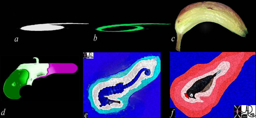 The shape of the pancreas is quite unusual in biology and has been described as a comma on its side (a), a 9 or 6 on its side (b) a bananna (c) an old fashioned revolver (d), a seahorse (e) and a woodpecker (f) 41502 Courtesy Ashley Davidoff MD
We will devote some time to the description, because subtle change in the shape may reflect important underlying disease. Identification of this alteration can mean the difference between making an early diagnosis of pancreatic carcinoma and saving a patient from a horrible disease, or missing the diagnosis and losing the opportunity. Adenocarcinoma of the pancreas is usually a rapidly fatal disease but the few who are fortunate to survive the illness have had the diagnosis made early, when surgical resection was feasible. It is imperative that routine assessment of the pancreas should focus on subtle shape and textural changes.
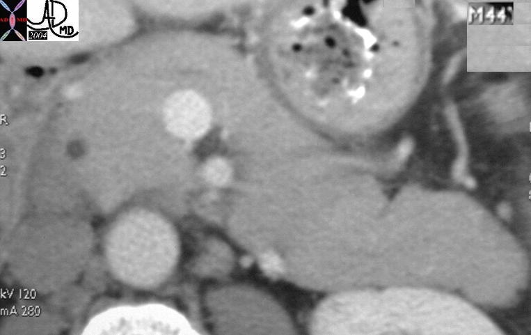 44 year old male with normal appearing pancreas. Note relatively smooth border 25034 Courtesy Ashley Davidoff MD pancreas normal anatomy imaging radiology CTscan time age aging growth
Shape of the Head Of The Pancreas
The head has the most complex shape. It is generally considered to be globular as viewed in the coronal and A-P projection, while it is almost tear drop in shape when viewed in the sagittal plane. It is the medial and lateral extensions of the head that contribute to it’s complexity.
The uncinate process, is the medial extent of the head and true to the origin of the word, is hook shaped. In latin – uncus means “hook”. It shares its latin origins and shape with at least six other structures in the body, including the uncus of the hippocampal region in the brain, as well as multiple bones including the ethmoid, cervical vertebra, scapula, hamate, and third phalanx of the foot.
|
The lateral extent of the head represents the embryonic ventral portion responsible for housing the bile duct, duct of Wirsung and the ampulla. It is in intimate contact with its embryological brother the duodenum. The shape of this ventral portion at the point of contact with the duodenum varies from being almost flat and rectangular to a lobster claw shape. This portion of the pancreas usually occupies about 25% percent of the circumference of the duodenum. In the congenital anomaly called annular pancreas, the ventral portion forms a ring around the pancreas occupying the entire circumference usually causing varying degrees of duodenal obstruction.
Shape of the Neck of the Pancreas
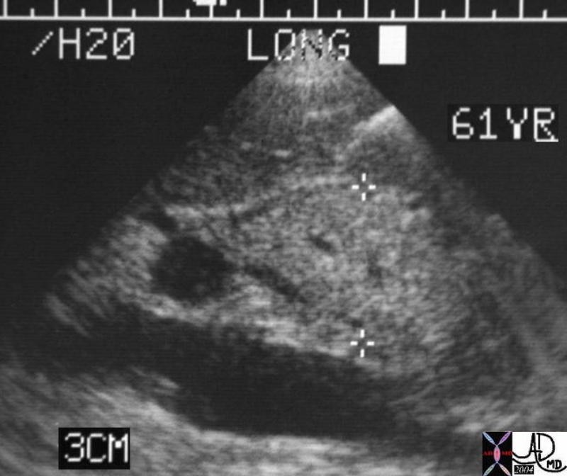
This series of six images show a normal pancreas from superior to inferior. The tail being the most superior first comes into view, and then the body, head and finally the uncinate process. The duct is seen as a faint lucency in c. Courtesy Ashley Davidoff MD 38031c01 code pancreas normal code pancreas normal anatomy Shape of the Body of the Pancreas The body and tail of the pancreas are less complex and form an elongated and irregularly prismatic structure. In cross section the body and tail have an anterior, posterior, and inferior surface making up the three borders of the 3 sided prism. This prismatic shape is best appreciated on a longitudinal view of the pancreas using ultrasound. The superior aspect of the pancreas is not usually appreciated on axial imaging due to its narrowed format and the intimate association with the splenic artery which partial volumes the interface. At the neck body interface anteriorly, a small protrusion called the omental tuberosity, arises from its superior and anterior border, just inferior to the origin of the celiac axis.
Shape of the Tail The tail is a continuation of the 3 sided prism representing its apex. It is usually narrowed and thinned and sometimes almost pointed at the left extremity. In the young it is more commonly is thickened, flattened or bifid.
Shape of the Surface Of The Gland The surface of the pancreas is relatively smooth in the young but as the gland starts to age and involute, the interstitium is replaced by fat resulting in a nodular surface. This finding is more apparent in the body and tail than in the head.
The pancreas is a soft organ and it’s form can be altered by the structures surrounding it. This particular pattern is exemplified by the patient who has had a left nephrectomy in whom the pancreas will flop back to take up the empty space. The angle between the neck and body thus becomes almost 90 degrees . Other parts may be pushed or pulled depending on the amount of retroperitoneal fat that is present. In patients with large amount of fat, the “push-pull” effect would be cushioned and lessened, while the reverse would be true in patients with little fat to pad the effect. The push effect may be seen in patients with abdominal aortic aneurysms where the aneurysm displaces the pancreas anteriorly. The right kidney may push the junction of the body and tail forward while the remaining portions of the tail curls around the kidney in sinuous fashion. Even the fluid filled stomach seems to be able to push the pancreas and deform it. Sometimes even a stomach filled with fluid seems to have the capacity to push the pancreas around. Thus the pancreas seems to be a “softy” and is pushed around, but, as many experienced surgeons tell their residents – “don’t mess with pancreas” Disruption of this very soft organ leads to chemical warfare in the body that create a far reaching destruction, and sometimes death !!!! Shape – Application to Disease and Imaging The shape of the pancreas as noted above, has wide variation depending on the part of the pancreas under consideration, age of the patient, body habitus, normal and abnormal structures surrounding the pancreas. In general it is reasonable to divide the pancreas in two groups in the evaluation; the neck body and tail as one entity, and the head uncinate process and ventral remnant as the second entity. The reason for doing this is that neck body and tail can usually be evaluated as one entity over a short craniocaudad distance and usually have similar texture and character. The head is usually the most inferior part, lies in a different axial plane, and is slightly more complex in its structural makeup. It has a wide range of variability and has the added component of the bile duct and ampulla to consider. Probably the most important and most difficult challenge in pancreatic evaluation is the need to identify an early asymptomatic carcinoma of the pancreas. Shape deformity and aberrance in enhancement pattern are the key issues to keep in focus. The ductal acinar carcinoma, represents the most common form of carcinoma. Its most important characteristic is that it has poor blood supply and presents as a hypovascular mass. In the body and tail it is not difficult to identify, but in the head and uncinate process the “normal heterogeneity of the head, created by the distal bile duct, downstream pancreatic ducts, and a limited space where so many structures are converging. Meticulous attention to technique with a contrast filled and distended duodenum, optimal vascular opacification and slice collimation, and compulsive observation is required to make an early diagnosis of this disease. The exophytic lesions are also important to focus on. Make sure in the evaluation that you evaluate beyond the immediate borders of the pancreas. There are tumors that have fooled the most experienced of observers being exophytic and missed. |
 The anterior and posterior edges of the hook in the pancreas should be straight or almost concave. Together with the globular head, the combination has a shape reminiscent of the head of a woodpecker with the uncinate process representing the beak. Masses growing in the uncinate process may change the biconcave beak shape into a more rounded shape. Pancreatitis both acute and chronic will result in the rounding out of the normal beak shape as well.39864b Courtesy Ashley Davidoff MD code pancreas
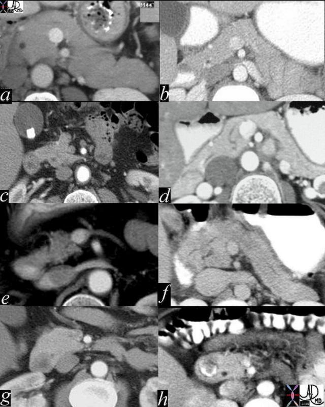 The size and shape of the head varies considerably among individuals. The series of CT scans therough the head and uncinate process shows a small compact head in a, thin elongated head with a prominent reversed c shape to the ventral portion which is in contact with the duodenum. (b). A variety of normal shapes and sizes of the ventral portion is seen in c,d,e,and f. The position of the duct in f suggests a pancreas divisum. Partial annular pancreas is noted in g, while a full annular pancreas without obstruction is seen in h.41502c02 Courtesy Ashley Davidoff MD
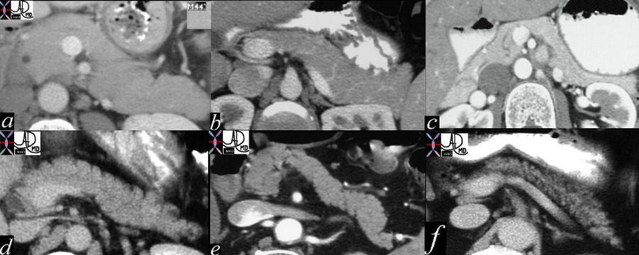 The variations of the size and surface shape of the body are noted in the above cases with the full and smooth body of youth in a, the gracile and smooth bodies of youth in b and c. Aging brings on atrophy and a wrinkling as evidenced by nodularity with a variety of manifestations. In d, the anterior border shows nodularity caused by early infiltration of fat , while in e atrophy is manifest with nodularity on both sides associated with thinning of the gland. In the last case (f) the nodularity and thinning caused by the fat infiltration is marked. 41505c02 Courtesy Ashley Davidoff MD
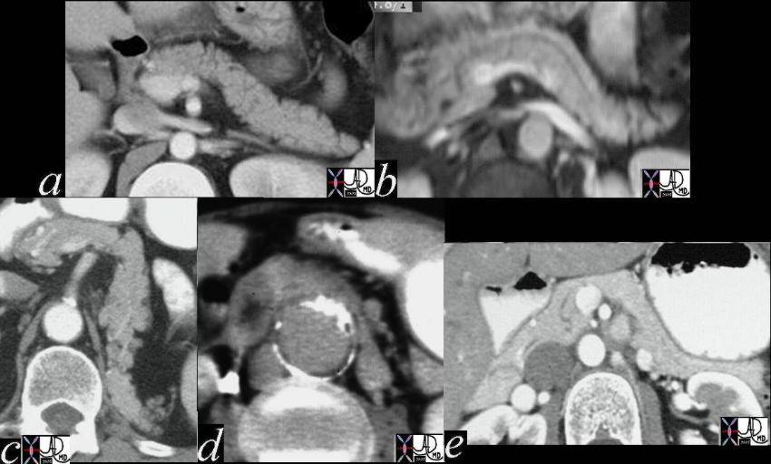 The shape and body of the tail varies between individuals sometimes as an innate shape of th structure itself and sometimes by the push and pull of the neighboring structures. The body is relatively straight in a, mildly sigmoid shaped in b, pulled by the absence of the kidney to look like a “7” caused by absent kidney, pushed from behind by an aneurysmal aorta in d, and impinged upon by the stomach in e. 41505c03 Courtesy Ashley Davidoff MD code
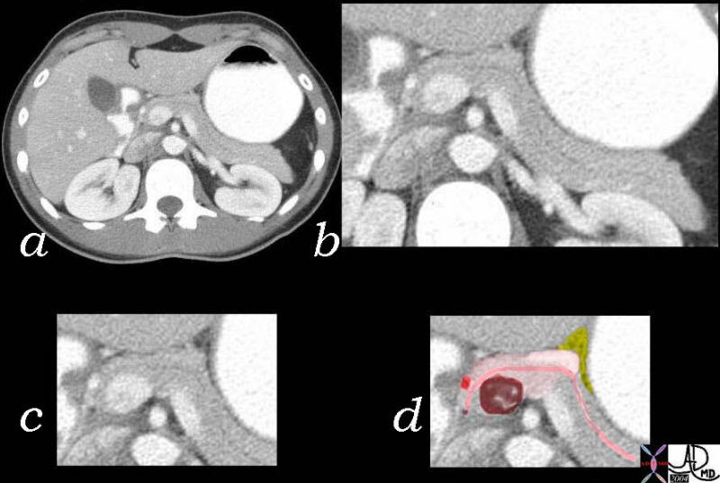 This series of CT images shows the neck of the pancreas (light pink) limited on the right by the gastroduodenal artery, (red) lying inf front of the SMV. Its left side has no clearly defined border but at the junction of neck and body there is sometimes a protuberance called the tuber omentale which is seen in this case outlined in solid pink in image d. The lesser omentum (yellow) is in contact with the tuber omentale. The pancreatic duct is well seen and normal in this case (dark pink) 38025c003 Courtesy Ashley Davidoff MD38025c003 Courtesy Ashley Davidoff MD