
Imaging the Pancreas – using MRI as a tool
Author Ashley Davidoff MD
Collaborators Charles Allison MD Adam Asarch MSII David Lee MD Scott Tsai MD Sam Yam PhD.
FINDINGS ON MRI
Using MRI for Masses
Using MRI for Calcifications
Using MRI for Cysts
Using MRI for Ductal Disease
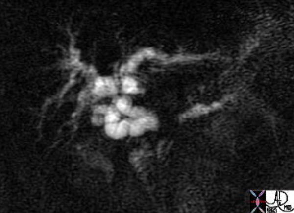 The image from an MRCP shows the MRI version of the double duct. The separation of the two ducts suggests a large size of the mass. This patient had pancreatic carcinoma. 41371 Courtesy of Ashley Davidoff MD
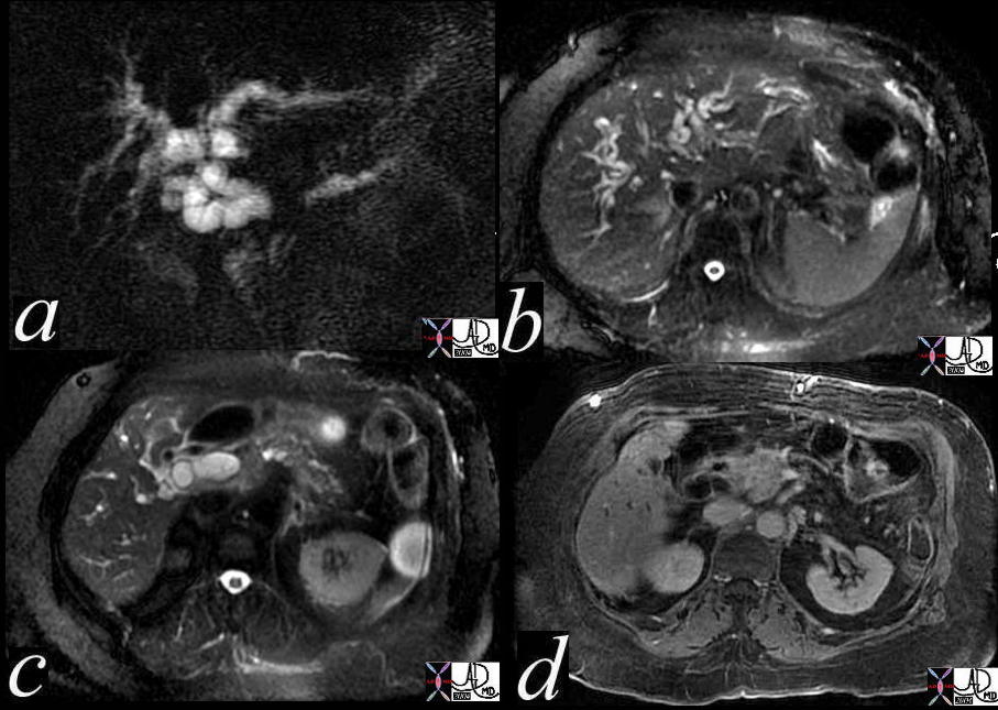 The image from an MRCP shows the MRI version of the “double duct” sign. The separation of the two ducts suggests a large size of the mass (a). Image b shows intrahepatic biliary dilatation, while image c shows CBD dilatation. Image d shows a mass in the head of the pancreas on the contrast enhanced sequence. This patient had pancreatic carcinoma. 41371c Courtesy of Ashley Davidoff MD
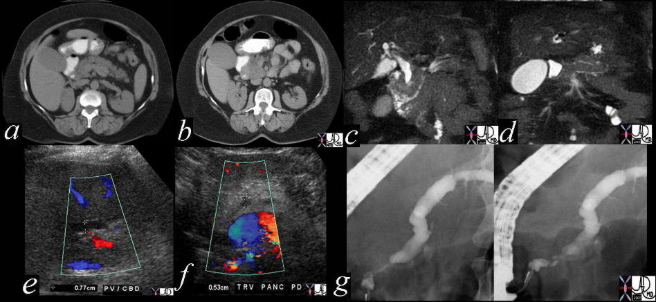 This combination studies includes CTscans, MRIs, US scans, and an ERCP. They suggest a subtle mass in the head of the pancreas with early bile duct dilatation and more obvious pancreatic duct involvement. This was a case of adenocarcinoma of the head of the pancreas. 41293a16c Courtesy Ashley Davidoff MD
Using MRI for Fat
DISEASES ON MRI
Using MRI for Acute Pancreatitis
Using MRI for Carcinoma
The image from an MRCP shows the MRI version of the double duct. The separation of the two ducts suggests a large size of the mass. This patient had pancreatic carcinoma. 41371 Courtesy of Ashley Davidoff MD
The image from an MRCP shows the MRI version of the “double duct” sign. The separation of the two ducts suggests a large size of the mass (a). Image b shows intrahepatic biliary dilatation, while image c shows CBD dilatation. Image d shows a mass in the head of the pancreas on the contrast enhanced sequence. This patient had pancreatic carcinoma. 41371c Courtesy of Ashley Davidoff MD
This combination studies includes CTscans, MRIs, US scans, and an ERCP. They suggest a subtle mass in the head of the pancreas with early bile duct dilatation and more obvious pancreatic duct involvement. This was a case of adenocarcinoma of the head of the pancreas. 41293a16c Courtesy Ashley Davidoff MD
Using MRI for Cysts
Using MRI for Pancreas Divisum
Using MRI for Serous Cystadenoma
Using MRI for Mucinous Cystadenoma
Using MRI for Annular Pancreas