
BLOOD SUPPLY
The pancreas has a very rich blood supply from both the celiac axis and the superior mesenteric artery (SMA). Typically (in 90% of people), the celiac axis divides into the common hepatic, splenic, and left gastric arteries. The common hepatic artery gives rise gastroduodenal artery (GDA), and then turns upward to the porta hepatis. The GDA courses from its origin behind the first part of the duodenum and, after giving off the superior pancreaticoduodenal artery, lies on the anterior surface of the head of the pancreas. At the lower border of the first part of the duodenum, it branches into the right gastroepiploic artery and the superior pancreaticoduodenal artery.
The superior pancreaticoduodenal artery gives off two major branches – the anterior pancreaticoduodenal and the posterior pancreaticoduodenal artery. These two latter vessels form an arcade around the head of the pancreas and reform inferiorly as the inferior pancreaticoduodenal artery. The inferior pancreaticoduoudenal artery is a branch of the SMA.
For the most part, the head of the pancreas and the duodenum have a common blood supply: the anterior and posterior pancreaticoduodenal arcades.
The body and tail receive blood from both the splenic artery and from the SMA as well.
Blood Supply from the Celiac Axis
 Celiac Axis Celiac Axis |
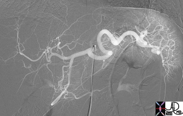
| In this digitally subtracted angiogram of the celiac axis, the main branches and secondary branches of the pancreaas are exemplified. Can you identify them?
39762 Courtesy Ashley Davidoff MD |
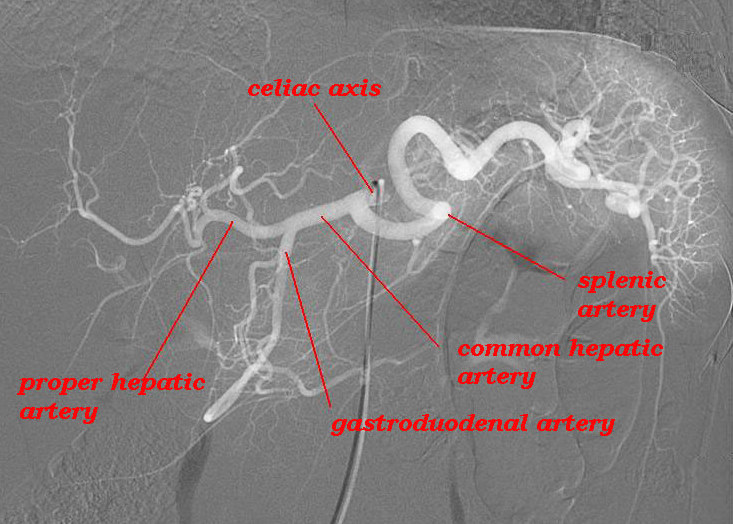 Celiac Axis Celiac Axis |
| The digital angiogram shows the major branches of the celiac axis which is the artery of the foregut. The celiac axis divides into two major vessels including the common hepatic artery and the splenic artery. The left gastric artery also usually arises from the celiac axis, but is not seen in this study. The two major branches of the common hepatic artery are the gastroduodenal artery (GDA) and the proper hepatic artery.. The unlabelled secondary branches of the splenic artery include branches to the body and tail of the pancreas, the short gastric arteries to the stomach and the terminal branches to the spleen. The gastroduodenal artery supplies branches to the pancreatic head, the duodenum, and the greater curvature of the stomach.
Courtesy Ashley Davidoff MD 39762L02 |
The celiac axis supplies the pancreas from both the common hepatic artery as well as the splenic artery. The splenic artery supplies 3 major branches to the body and tail. These include the dorsal pancreatic artery, (DPA) pancreatica magna (PM), and caudal pancreatic artery. (CPA)
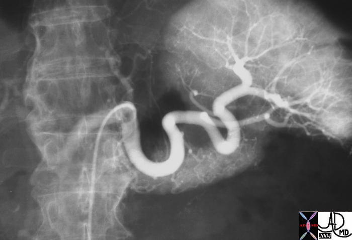
In this splenic artery angiogram the parenchymal blush of the pancreatic body and tail is well seen, but the individual branches are difficult to identify.
39842b Courtesy Ashley Davidoff MD
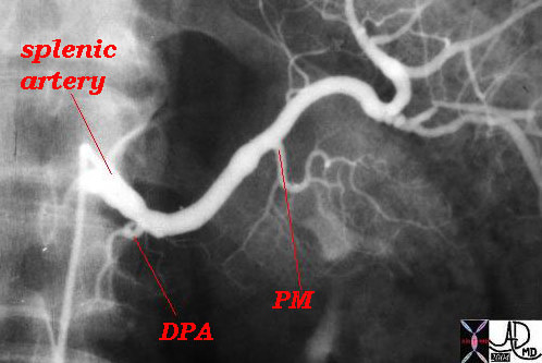 Splenic artery branches to the pancreas Splenic artery branches to the pancreas |
| This angiogram demonstartes the first and second branches of the the pancreas arising from the splenic artery. The dorsal pancreatic artery (DPA) is the first and the pancreatica magna (PM) the second. In this case pancreatica magna does justice to its name and is a particulalrly large vessel in this patient. An early parenchymal blush canb be seen. The third branch, the caudal artery supplying the tail of the pancreas is not seen in this case.
Courtesy Ashley Davidoff MD 39762L01 |
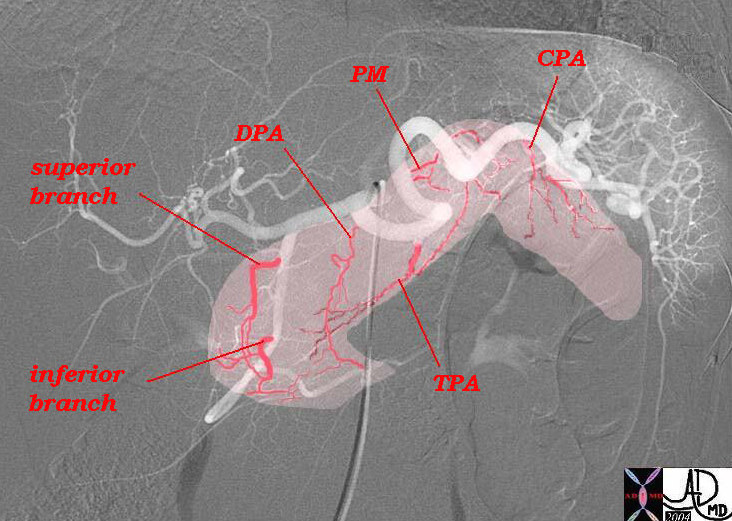 Supply of the pancreas Supply of the pancreas |
| In this digitally subtracted angiogram of the celiac axis the main branches and secondary branches of the pancreas are exemplified. There are three major branches to the body and tail of the pancreas. The first branch off the splenic artery is the dorsal pancreatic artery (DPA) which splits as an inverted “T” to form the transverse pancreatic artery. (TPA) Next branch up is the pancreatica magna (PM) which also contributes to the transverse pancreatic artery (TPA) while the last branch in the tail is called the caudal branch of the pancreatic artery. (CPA) The first branch off the superior aspect of the GDA is the superior pancreaticoduodenal artery which communicates with the inferior pancreaticoduodenal artery which in this instance also arises from the GDA . These two vessels connect in the anterior and posterior pancreaticodudenal arcades.
Courtesy Ashley Davidoff MD 39762b03b01L02 |
Blood Supply from the SMA
The SMA contributes to the blood supply of the head of the pancreas as well the tail. The head receives a branch from the SMA called the inferior pancreaticoduodenal artery which divides into anterior and posterior branches each of which connects to the anterior and posterior branches of the superior pancreatic branch from the GDA (see above diagram). The head of the pancreas is thus well supplied with blood in the form of a pancreaticoduodenal arcade.
The Pancreaticoduodenal Arcade
This arcade has both an anterior component and a posterior component. The anterior component is formed by the anastomosis of the anterior branch of the superior pancreaticoduodenal artery, which is a branch of the gastroduodenal artery, and the anterior inferior pancreaticoduodenal artery, which is usually a branch of the superior mesenteric artery.
The posterior pancreaticoduodenal arcade is formed by the anastomosis of the posterior branch of the superior pancreaticoduodenal artery, usually a branch of the gastroduodenal artery, and the posterior branch of the inferior pancreaticoduodenal artery, which usually arises from the SMA. The posterior branch of the superior pancreaticoduodenal artery courses to the right and anterior to the common bile duct, and provides blood supply to the extrahepatic duct. During sphicnterotomy this vessel can be severed and be a major source of bleeding.
The duodenum and head of the pancreas share their blood supply and the duodenum can be comprmised by total pancreatectomy. Additionally if a patient requires embolisation for a pseudoaneurysm in the pancreatic head (a rare complication of pancreatitis), embolisation must be approached from both the celiac axis and the SMA because of the dual and connecting blood supply.
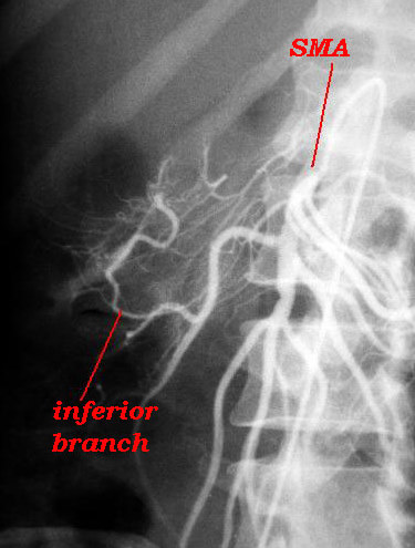
Blood supply to the head of the pancreas from the SMA |
| In this injection of the SMA the pancreaticoduodenal arcade is well demonstrated. The inferior pancreaticoduodenal artery arises from the SMA and gives off branches to the duodenum and the pancreas.
Courtesy Ashley Davidoff MD 39797L01 |
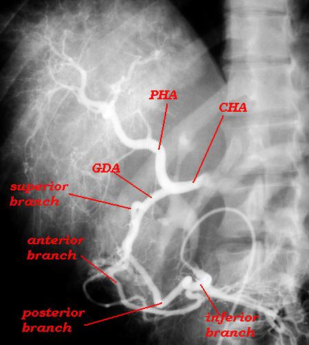 Pancreaticoduodenal arcade Pancreaticoduodenal arcade |
| In this angiogram the SMA not only fills the pancreatic branches to the head of the pancreas but also the GDA and subsequently the CHA due to a stenosis of the celiac axids and retrograde flow. The inferior and superior branches both give rise to anterior and postrerior divisions which connect and form the pancreaticoduodenal arcade. The branching pattern in the liver is heterogeneous particulalrly at the inferior edge of the liver. These finding represent diffuse infiltrative metastases from gastric carcinoma.
Courtesy Ashley Davidoff MD 39821L02 |
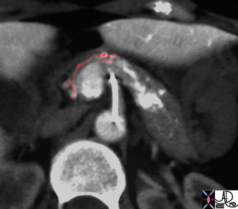 SMA contribution to the head SMA contribution to the head |
| This is an arterial phase of a CT portogram using direct arterial injection into the SMA via a catheter in the SMA. The branches to the head via the anterior component of the pancreaticoduodenal arcade can be seen anterior to the head and neck of the pancreas. (red overlay) Courtesy Ashley Davidoff MD 39849b03 |
 SMA contribution to the body – transverse pancreatic artery SMA contribution to the body – transverse pancreatic artery |
| This is an arterial phase of a CTscan through the body of the pancreas and a branch with a “T” shaped distribution is seen arising from a branch of the SMA, representing the transverse pancreatic artery. The vessel just posterior to the neck of the pancreas which gives rise to the transverse pancreatic artery is the posterior inferior pancreaticoduodenal artery – a branch of the SMA.
Courtesy Ashley Davidoff MD. 25226 L02 |
 SMA contribution to the body – transverse pancreatic artery SMA contribution to the body – transverse pancreatic artery |
| In this CT portogram the injection of contrast was into the SMA and the perfusion to the the body of the pancreas with a portion of the medial part of the head of the pancreas also showis some contrast perfusion. The lateral part of the head does not perfuse with this injection suggesting that is fed mostly by tjhe celiac axis in this case. 39850b01 Courtesy Ashley Davidoff MD |
LYMPHATIC DRAINAGE
The lymphatic channels originate around the acini and initally following the capillaries and subsequently following the arterioles and arteries. No lymphatics are found around the islets of Langerhans. The lymphatics of the pancreas follow the branches of the celiac axis and the SMA that supply the pancreas, with the nodal supply of the head following the hepatic artery and the supply of the body and tail following the splenic artery. The final regional pathway for the celiac nodes and the SMA nodes will be to the paraortic nodes.
The nodes of the hepatic vessels completely surround the head, and are found anterior, posterior, superior, and inferior to the head. The head of the pancreas shares these nodes with the duodenum with which it is intimate contact.
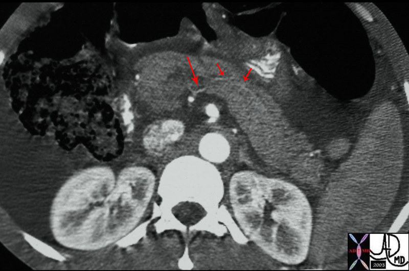
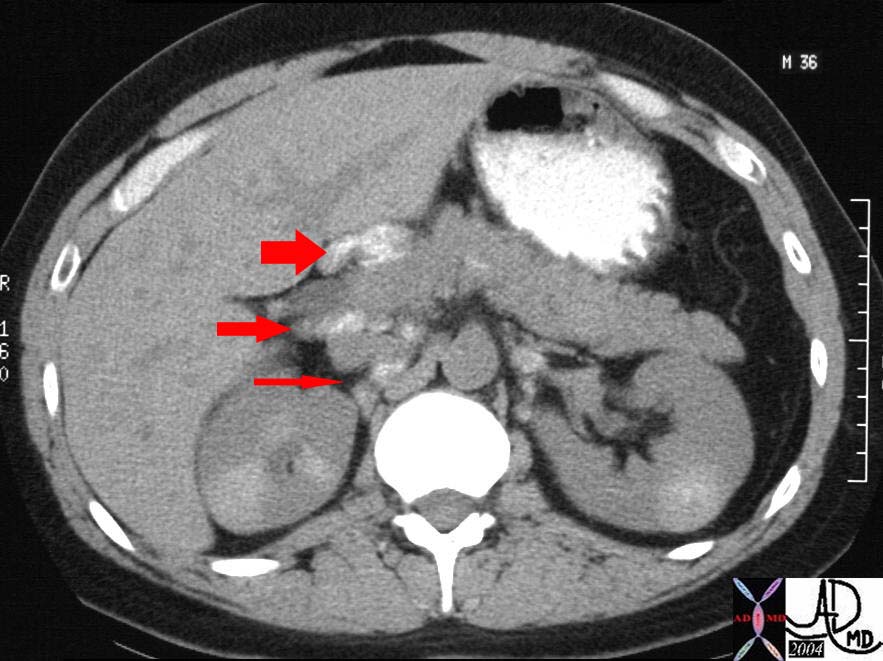 Lymph nodes of the head of the pancreas Lymph nodes of the head of the pancreas |
| The CT through the mid abdomen and kidneys is from a patient with sarcoidosis in whom there is extensive but fine calcifications in the kidneys and the celiac axis lymph nodes surrounding the pancreas. These are abnormal lymph nodes but the case serves to demonstrate the lymphatic associations of the head of the pancreas. There are nodes anterior to the head in the porta hepatis (fat arrow), and in the portocaval space posterior to the head. (middle arrow) More posteriorly (thin arrow) small calcified nodes are seen between the IVC and the right crus of the diaphragm.
Courtesy Ashley Davidoff MD 26477b02arrow |
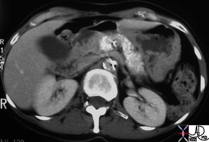
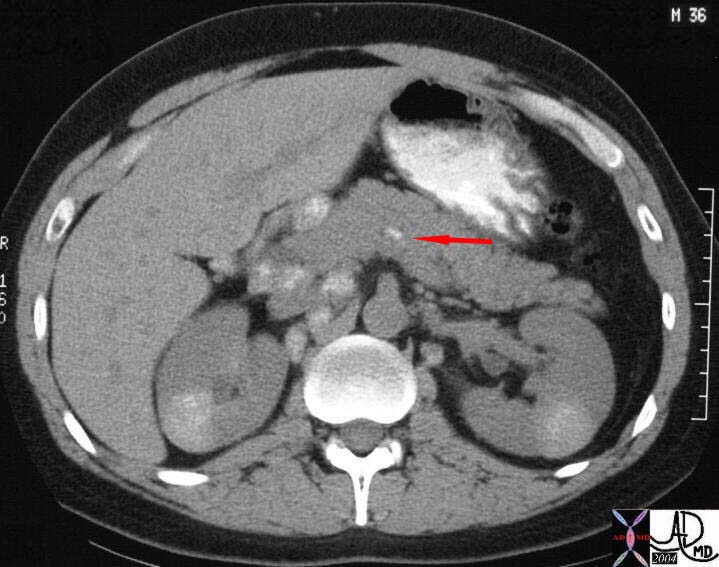 Intrapancreatic lymph node Intrapancreatic lymph node |
| In this CT of the same 36 year old patient with sarcoidois a small calcified intrapancreatic node is seen at the junction of the neck and body of the pancreas. There are also nodes anterior and posterior to the head in the porta hepatis and more posteriorly small calcified nodes are seen between the IVC, the right crus of the diaphragm and the right kidney.
Courtesy Ashley Davidoff MD 26477b03arrow |