
Author Ashley Davidoff MD
Collaborators Charles Allison MD Adam Asarch MSII David Lee MD Scott Tsai MD Sam Yam PhD.
Approach
The pancreas shows unusual changes over time. In the young adult, and particularly in the female, the tail often appears enlarged while the head of the pancreas appears relatively small. With aging the tail becomes less prominent while the head becomes relatively large. The most common cause of enlargement of the pancreas is pancreatitis. Because of the absence of a capsule, inflammatory fluid leaks from the pancreas into retroperitoneum and so the enlargement of the pancreas itself may be only minimal, even in severe pancreatitis. Classical pancreatic scirrhous carcinoma of the pancreas usually presents with a focal relatively small hypovascular mass while the islet cell tumors can be larger and are hypervascular. Lymph nodes surround the pancreas, and when they become large and confluent in lymphoma and metastatic disease they merge imperceptibly or invade the pancreatic tissue. This results in a diffuse enlargement of the pancreas. Sometimes the cystic duct becomes so enlarged that it looks like the pancreas and a cystic enlargement must raise the possibility of this entity.
Differential Diagnosis
Normal variant
Pancreatitis and its complications
Dilated pancreatic duct
Malignant tumors especially islet cell tumors
Metastatic disease (lymphoma)
Von Hippel-Lindau
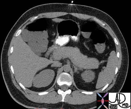 This CTscan of a young adult with abdominal discomfort shows a relatively enlarged body and tail of the pancreas. This is a normal finding. In the young the body and tail tend to be relatively large and in the elderly the reverse is true. 40770b01 Courtesy Ashley Davidoff MD
 This CTscan of a 35 year old female with epigastric discomfort with abdominal pain shows a relatively enlarged body and tail of the pancreas. This is a normal finding. In the young the body and tail tend to be relatively large and in the elderly the reverse is true. 41394
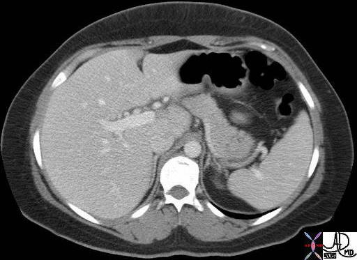
 This a CTscan through the head neck and body of a normal pancreas in a aging adult. It shows the relative enlargement of the head of the pancreas and relatively small size of the body. This is a normal finding. 19387 Courtesy Ashley Davidoff MD
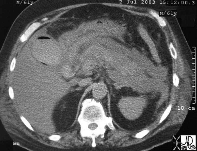
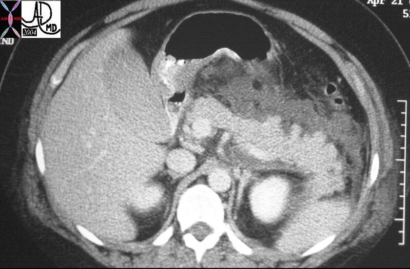
This is a CTscan of a 61 year old man who presents with abdominal pain, acute inferior myocardial infarction, with a background history of type II diabetes. The overall size of the pancreatic bed has increased mostly due to the peripancreatic effusion. The pancreas itself is only minimally enlarged and has a subtle increase in density suggesting hemorrhagic pancreatitis. It is sandwiched between the exudates anteriorly and posteriorly. Note the air in the lumen and the wall of the gallbladder consistent with emphysematous cholecystitis. Note also the changes in the left lateral conal fascia, and properitoneal fat suggesting transgression of fluid. Within both the hypodermis of both flanks there are soft tissue changes consistent with radiologic Gray-Turners sign. 31093 Courtesy Ashley Davidoff MD
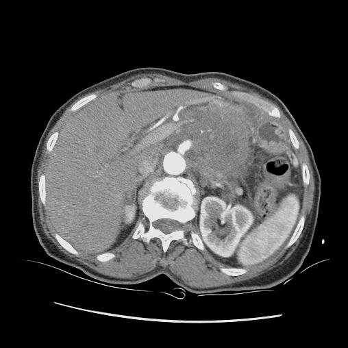
 This CT scan through the pancreas shows a relatively normal sized pancreas with a large peripancreatic effusion. The absence of a pancreatic capsule allows free leakage of the exudative component of the inflammation out of the pancreas and into the anterior pararenal space. 15730 Courtesy Ashley Davidoff MD
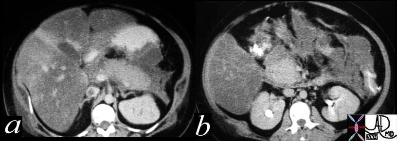 This combination CTscan shows the manifestations of severe acute alcoholic pancreatitis with mild enlargement of the pancreas but a significant peripancreatic exudate. The exudate extends from the anterior pararenal space to the peritoneal cavity and into base of the mesentery. Associated findings include IVC thrombosis, and impingement onto the greater curvature of the stomach. Severe steatosis of the liver with areas of sparing is also noted. 40277c Courtesy Ashley Davidoff MD
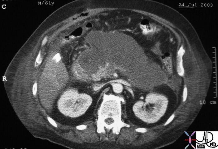
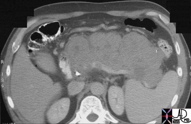
The CT scan of the abdomen of a 61 year old male shows a large cystic type lesion in the lesser sac, conforming to the shape of the pancreas. Note also the piecemeal appearance of the remaining viable pancreas in the region of the uncinate process. There was little other viable pancreas identified and a diagnosis of pancreatic necrosis was made. Associated findings include extension of the inflammatory process to the left paracolic gutter and through to the soft tissues of the flanks, as well as postoperative air in the right rectus abdominis muscle. 31188b01 Courtesy Ashley Davidoff MD
 This CT scan through the pancreas shows a mass in the body of the pancreas associated with encasement of the portal vein and splenic artery ((subtotal occlusion) with an almost complete encirclement of the celiac axis by a necklace of aggressive tissue. These findings are consistent with a diagnosis of a primary carcinoma of the pancreas. Based on the desmoplastic nature of the process the tumor is most likely an adenocarcinoma. Courtesy Ashley Davidoff MD18103
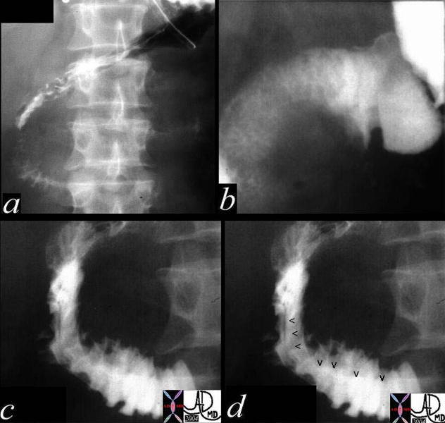 This upper GI study is of a young male who sustained blunt trauma to the abdomen and was complaining of abdominal pain. The images show the C-sweep of the duodenum which is widened, with extrinsic mass effect (arrowheads in d). The trauma to the abdomen was complicated by traumatic pancreatitis 18566c04 Courtesy Ashley Davidoff MD
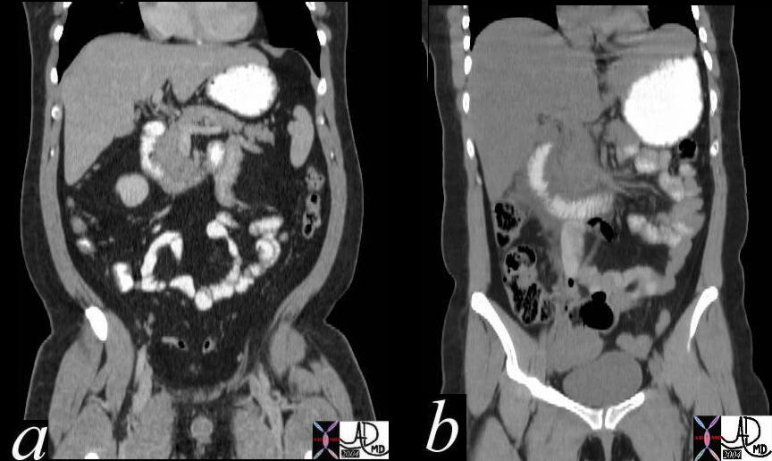 This combination of coronally reformatted images of the abdomen show a normal c sweep in a, and an enlarged and widened c-sweep in b. The patient in b, had acute pancreatitis with swelling of the pancreatic head caused mostly by the exudate which can be seen on either side of the duodenum. 40999c02 Courtesy Ashley Davidoff MD
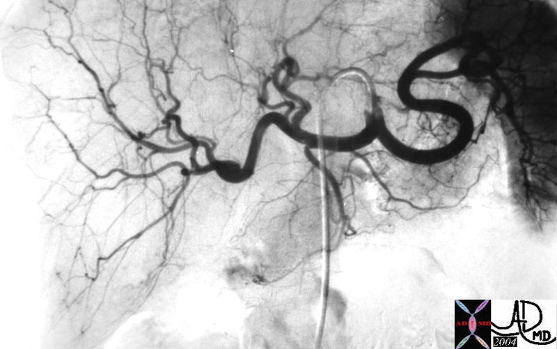 This angiogram is from a 50 year old female with acute abdominal pain with elevated amylase and a diagnosis of acute pancreatitis. The pancreas is mildly enlarged 40918 Courtesy Laura Feldman MD
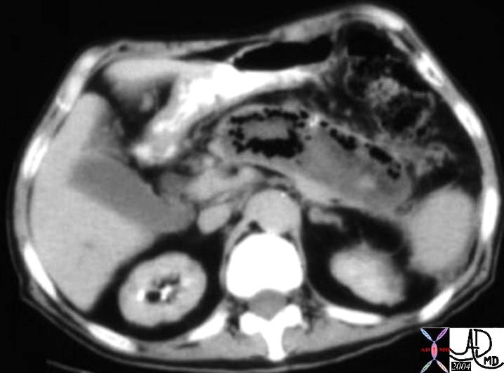 This is a CT of an elderly man who had severe pancreatitis. Within the pancreatic bed there is a structure that conforms to the shape of the pancreas. It is enlarged and consists mostly of air, fluid, and high density hemorrhagic components. The findings are consistent with a gangrenous pancreatic abscess. The surgeon noted that the pancreas looked and smelled like a “dead fish…. that had been dead for quite a long time.” 04959 Courtesy Ashley Davidoff MD
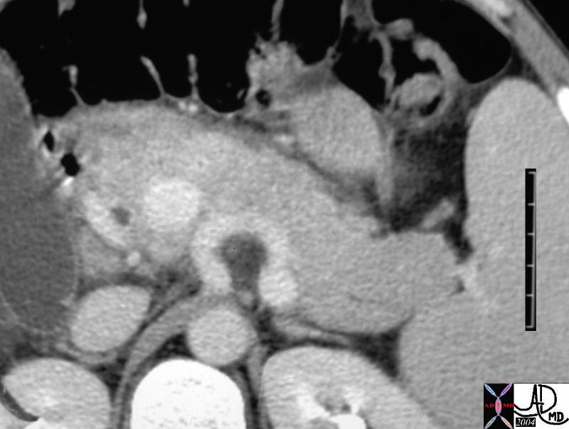 This coned down view of the CTscan of the pancreas of a young man with abdominal pain the tail of the pancreas is relatively large and the head relatively small consistent with the patients age. However on closer inspection there is a thin line of exudative fluid anterior to the pancreas in the anterior pararenal space. He was known to have both histoplasmosis and MAI and clinically his biochemistry was consistent with an infectious pancreatitis, most likely of MAI origin. 40092b01 Courtesy Ashley Davidoff MD
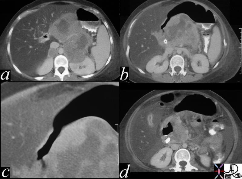
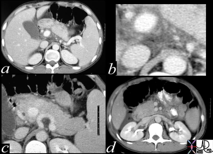 Image c in this combination series of the CTscan of the pancreas of a young man with abdominal pain, shows prominence to the tail of the pancreas and a relatively large and the head relatively small consistent with the patients age. However on closer inspection there is a thin line of exudative fluid anterior to the pancreas in the anterior pararenal space. In a, there is fluid posterior to the pancreas, better seen in b, and there is fluid in Morrison’s pouch (d). He was known to have both histoplasmosis and MAI and clinically his biochemistry was consistent with an infectious pancreatitis, most likely of MAI origin. 40092b01 Courtesy Ashley Davidoff MD
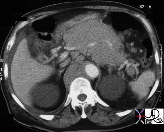 The CTscan in this 87 year old male is through the body of pancreas and shows, homogeneous deformity and enlargement of the pancreas. Associated findings include small retropancreatic nodes, with larger retrocrural nodes and ascites. The findings are consistent with a diagnosis of lymphoma. Incidental findings include bilateral upper pole renal cysts. 29945 Courtesy Ashley Davidoff MD
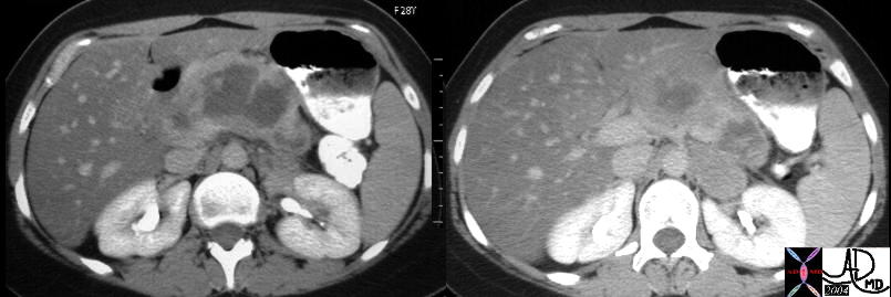 The CTscan in this 28 year old female shows an enlarged body of the pancreas, with a matrix which is hypodense relative to the thick periphery. She has a known diagnosis of non Hodgkin’s B cell lymphoma. Associated findings include diffuse severe steatosis of the liver. 40425c01 Courtesy Ashley Davidoff MD
 This combination CTscan series reveals an enlarged heterogeneous pancreas in an adult female patient with a known abdominal mass (a). A bile duct stent is noted in b, with extrinsic compression, narrowing of the gastric antrum noted in c and d. At endoscopy invasion of the stomach was identified. Associated severe fatty change of the liver is present. This is a case of pancreatic lymphoma. 40859c Courtesy Ashley Davidoff MD
 This CTscan through the body of the pancreas shows diffuse mostly homogeneous enlargement of the pancreas. There is a thrombus in the splenic vein and large venous collateral around the mass suggesting high grade splenic vein stenosis and impending obstruction. A bile duct stent implies impingement of the mass on the duct. There is a punctate radioopacity, possibly a calcification on the right anterior border of the mass. This is a case of aggressive metastatic Merkel cell carcinoma to the pancreas 22322 Courtesy Ashley Davidoff MD
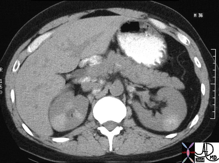
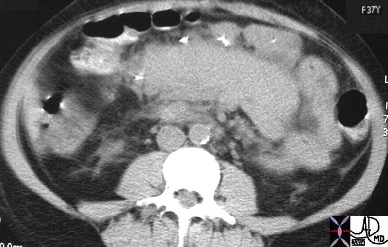 This CTscan shows homogeneous enlargement of the pancreas in a 37 year female with long standing diabetes and renal failure. Surgical clips of a recent pancreatic transplant are noted on the anterior border of the transplant. This is a case of pancreatitis of the transplanted pancreas. 25329 Courtesy Ashley Davidoff MD
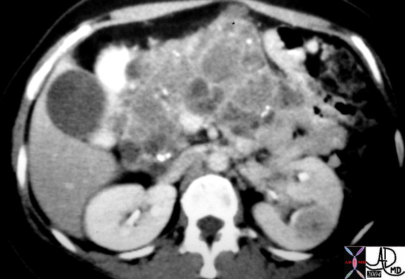 This CT scan through the pancreas shows diffuse heterogeneous enlargement of the pancreas, and a series of complex cystic and solid masses mass with punctate calcifications throughout the whole pancreas. Associated findings include a solid mass in the left kidney. These findings are consistent with a diagnosis of von Hippel-Lindau syndrome with a variety of cystic and solid, benign and malignant tumors of the pancreas 40531 Courtesy Ashley Davidoff MD
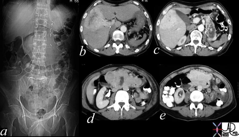 This is a CT combination series of abdominal images of a 66year old man with an abdominal mass for1 year. Image a, shows an epigastric mass pushing the transverse colon inferiorly. Image b shows a hypervascular metastasis in the liver, while c, d, and e show a diffusely enlarged pancreas replaced by a complex, heterogeneous mass that is mostly hypervascular. The findings of a slowly aggressive hypervascular neoplasm are not reminiscent of the classical adenocarcinoma of the pancreas. The histological type is not known in this case. 40834c Courtesy Ashley Davidoff MD
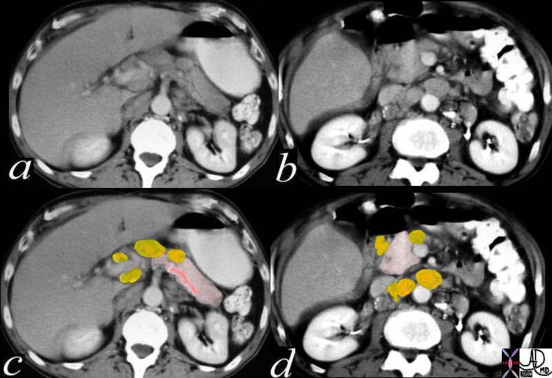 This combination CTscan reveals a slightly enlarged pancreas best appreciated in the body, neck head region (a). In this case discrete nodes are appreciated in the porta hepatis, portocaval space, periaortic region (yellow overlays in c and d) that account for the enlargement. This patient has a known diagnosis of Hodgkin’s lymphoma. 40432c02 Courtesy Ashley Davidoff MD
 This CT scan through the pancreas shows a series of calcified masses surrounding the head of the pancreas but also in the porta hepatis, portocaval space, and paracaval position. These findings represent lightly calcified lymph nodes. Associated findings include unusual wedge shaped calcifications in the left kidney, and mass like calcification in the right. This is a patient with sarcoidosis. The lymph node pattern in the foregut is not unusual for sarcoidosis. Kidney involvement with granulomatous disease is not uncommon, but the pattern of calcification in the kidney is quite unusual. Courtesy Ashley Davidoff MD 26477b02
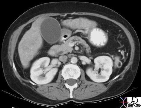
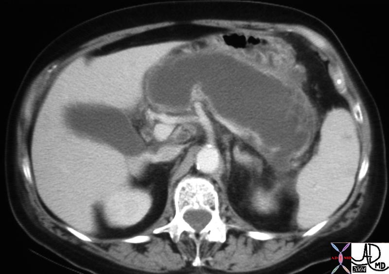 This CTscan through the pancreas shows a tubular cystic mass in the pancreatic body and tail, which conforms to the shape of the pancreatic tail and is associated with a thin rim of pancreatic tissue. The finding is consistent with an obstructed and dilated pancreatic duct with secondary pressure atrophy of the pancreas. The focal hypodensity in the spleen is of unknown significance. 40208 Courtesy Ashley Davidoff MD
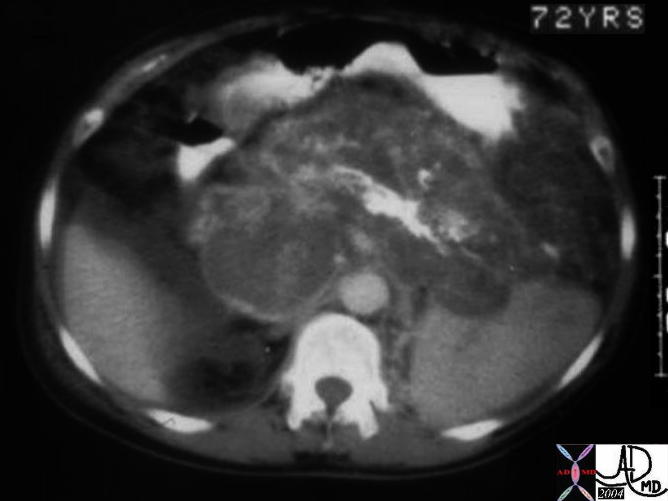 This CTscan of a 72 year old man through the body of an enlarged pancreas shows a central almost linear calcification with surrounding low density, representing the bulk of the parenchyma. Strands of soft tissue and blood vessels are scattered in the rest of the gland. At surgery this proved to be a giant hemangioma of the pancreas. This is not your usual case!! 04894c001 Courtesy Ashley Davidoff MD