
Imaging the Pancreas – using ERCP as a tool
Author Ashley Davidoff MD
Collaborators Charles Allison MD Adam Asarch MSII David Lee MD Scott Tsai MD Sam Yam PhD.
FINDINGS ON ERCP
Using ERCP for Masses
Using ERCP for Calcifications
Using ERCP for Cysts
Using ERCP for Ductal Disease
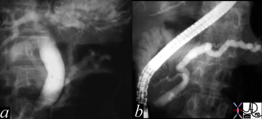 A 57 year old female presents with jaundice. Her ERCP shows dilatation of the bile duct and pancreatic duct – the “double duct” sign characteristic of adenocarcinoma of the head of the pancreas. In this case the stricture appears quite distal. Chronic pancreatitis can also rarely cause the “double duct” sign. In this case an ampullary carcinoma was the cause explaining the distal position of the strictures. 04655c03 Courtesy Ashley Davidoff MD
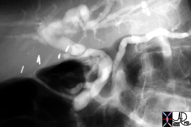 This is the injection of the MPD of the case above showing a small ventral duct of Wirsung. The case represents
pancreas divisum as seen by ERCP. The main duct was found opening separately within the pailla near the opening
of the CBD. 40617 Courtesy Ashley Davidoff MD
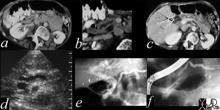 This combination series shows a dilated pancreatic duct in the head of the pancreas (a,b) as well as in the tail (c) The CBD is mildly dilated on US (d) The ERCP (e) shows that both ducts are dilated. At first glance a double duct sign seems likely since both ducts are dilated, but for different reasons. The main pancreatic duct drains into the accessory duct of Santorini, while the CBD drains in usual fashion into the ampulla. The smaller duct of Wirsung is seen mostly disconnected from the ductal system at large. This is a case of pancreas divisum. No pancreatic cancer was found on further workup 40617c01 Courtesy Ashley Davidoff MD
 This is an ERCP showing a “double duct” sign. With the heavy intraductal calcification chronic alcoholic pancreatitis is definite.
It is conceivable that the patient has a superimposed malignancy. In this case the double duct was caused by the strictures of
chronic pancreatitis. 41245c06 Courtesy Ashley Davidoff MD
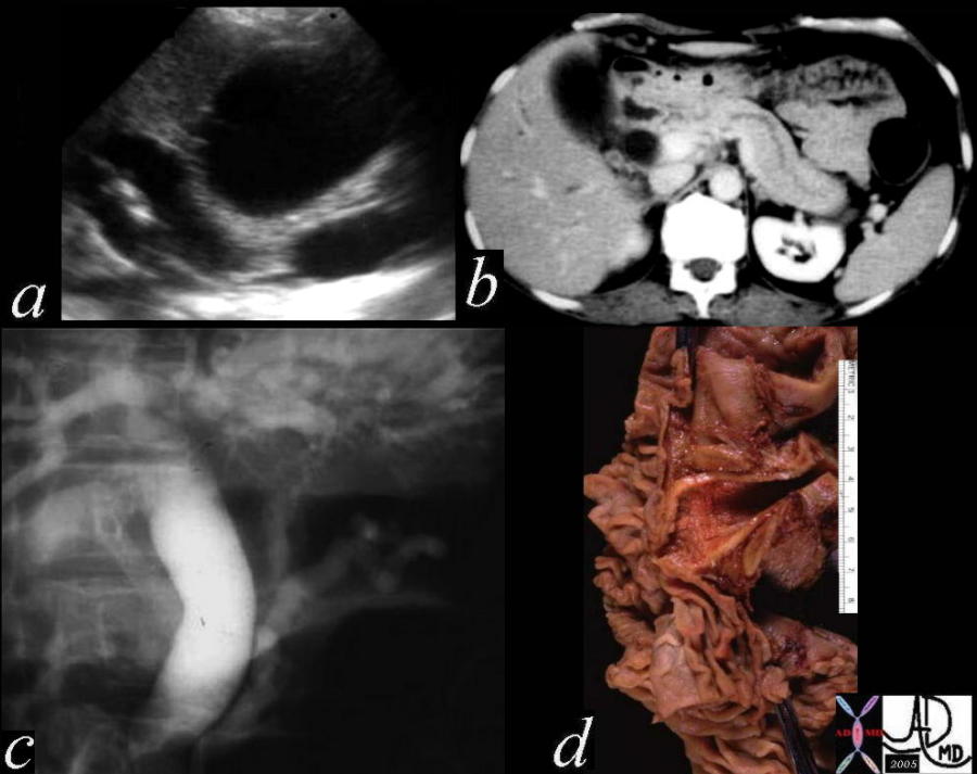
This combination studies includes CTscans, MRIs, US scans, and an ERCP. They suggest a subtle mass in the head of the pancreas with early bile duct dilatation and more obvious pancreatic duct involvement. This was a case of adenocarcinoma of the head of the pancreas. 41293a16c Courtesy Ashley Davidoff MD
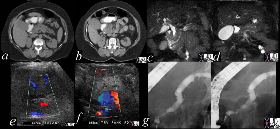
This is a series of images from the case history of a 57 year old female who presented with jaundice. The US image (a) shows a dilated bile duct and dilated pancreatic duct with no large mass between them. The CT shows a les impressive, but prominent pancreatic duct. The ERCP suggests a “double duct” sign. The pathology confirmed an ampullary carcinoma. 04655c05 Courtesy Ashley Davidoff MD
Using ERCP for Fat
DISEASES ON ERCP
Using ERCP for Acute Pancreatitis
Using ERCP for Chronic Pancreatitis
This is an ERCP showing a “double duct” sign. With the heavy
intraductal calcification chronic alcoholic pancreatitis is definite.
It is conceivable that the patient has a superimposed malignancy.
In this case the double duct was caused by the strictures of
chronic pancreatitis. 41245c06 Courtesy Ashley Davidoff MD
Using ERCP for Carcinoma
A 57 year old female presents with jaundice. Her ERCP shows dilatation of the bile duct and pancreatic duct – the “double duct” sign characteristic of adenocarcinoma of the head of the pancreas. In this case the stricture appears quite distal. Chronic pancreatitis can also rarely cause the “double duct” sign. In this case an ampullary carcinoma was the cause explaining the distal position of the strictures. 04655c03 Courtesy Ashley Davidoff MD
This combination studies includes CTscans, MRIs, US scans, and an ERCP. They suggest a subtle mass in the head of the pancreas with early bile duct dilatation and more obvious pancreatic duct involvement. This was a case of adenocarcinoma of the head of the pancreas. 41293a16c Courtesy Ashley Davidoff MD
Using ERCP for Cysts
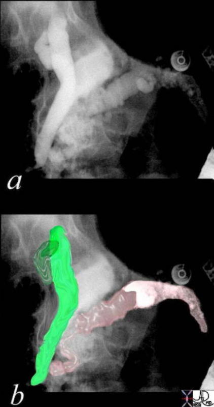
Using ERCP for Pancreas Divisum
This is the injection of the MPD of the case above showing a small ventral duct of Wirsung. The case represents
pancreas divisum as seen by ERCP. The main duct was found opening separately within the pailla near the opening
of the CBD. 40617 Courtesy Ashley Davidoff MD
This combination series shows a dilated pancreatic duct in the head of the pancreas (a,b) as well as in the tail (c) The CBD is mildly dilated on US (d) The ERCP (e) shows that both ducts are dilated. At first glance a double duct sign seems likely since both ducts are dilated, but for different reasons. The main pancreatic duct drains into the accessory duct of Santorini, while the CBD drains in usual fashion into the ampulla. The smaller duct of Wirsung is seen mostly disconnected from the ductal system at large. This is a case of pancreas divisum. No pancreatic cancer was found on further workup 40617c01 Courtesy Ashley Davidoff MD
Using ERCP for Serous Cystadenoma
Using ERCP for Mucinous Cystadenoma
Using ERCP for Annular Pancreas