
Imaging the Pancreas – using CT as a tool
Author Ashley Davidoff MD
Collaborators Charles Allison MD Adam Asarch MSII David Lee MD Scott Tsai MD Sam Yam PhD.
FINDINGS ON CT scan
Using CT scan for Masses
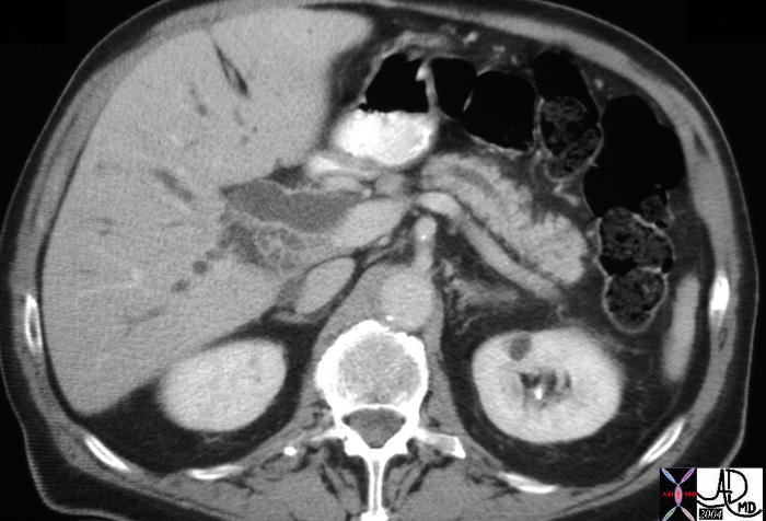
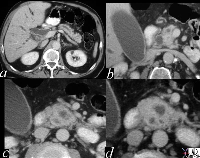 This CT scan series is of a patient with obstructive jaundice and shows a double duct sign suspicious for carcinoma off the head of the pancreas. A dilated pancreatic duct and a dilated CBD (“double duct” sign) is shown (a,b) and then 3 hypodensities and a rotund appearance of the head suggests an extra and unwanted component in the head of the pancreas. Can you identify the unwanted component from these images?. The findings nevertheless are consistent with a primary adenocarcinoma of the pancreas. 40840c01 Courtesy Ashley Davidoff MD
Using CT scan for Calcifications
 The CTscan through the body of the pancreas is of a 74 year old female with abdominal discomfort. She has no history of alcohol abuse and a normal US of the gallbladder. The study shows a complex, bilocular, cystic lesion in the tail of the pancreas with an enhancing rind, and two focal peripheral punctate calcifications. A diagnosis of a mucinous cystadenoma is thought to be most likely.
04811 Courtesy Ashley Davidoff MD
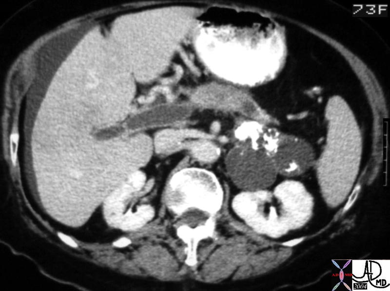 This CT scan through the pancreas shows a complex bicystic mass with dense peripheral curvilinear and dystrophic calcification in the tail of the pancreas. Associated findings include portal vein thrombosis (PVT) and ascites. These findings are consistent with a mucinous cystadenoma, and probable malignant transformation. 40712 Courtesy Ashley Davidoff MD
Using CT scan for Cysts
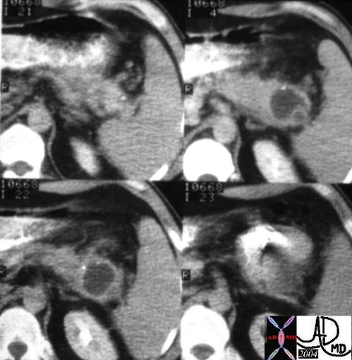
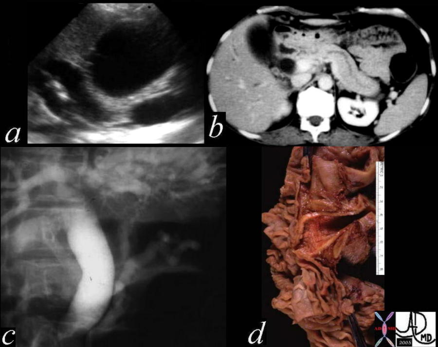
 This CTscan is through the tail of the pancreas shows what appears to be a simple cyst (a), but which proves to have a mural nodule on ultrasound (b). On the T1 weighted image (c) the nodule shows high intensity suggesting increased protein or acute blood, and the T2 (d) reveals the cystic nature of the matrix. At pathology the lesion was a benign serous cystadenoma. The high intensity on T1 was due to the high protein in the mucin. 40487c01 Courtesy Ashley Davidoff MD
Using CT scan for Ductal Disease
 This is a classical example of a double duct sign. The dilated pancreatic duct and bile duct almost point to the disease process in this image. A solid mass was identified in the head of the pancreas consistent with adenocarcinoma. 40837 Courtesy Ashley Davidoff MD
The combination of images below reflects the changes of the above case in at different levels and magnification.
 This is a classical example of a “double duct” sign. The dilated pancreatic duct and bile duct in a, almost point to the disease process in this image. The mass was as predicted to be in the head of the pancreas. In c and d, a relatively small hypodense mass was identified whose scirrhous nature, and critical location caused early obstructive jaundice. 40840c01 Courtesy Ashley Davidoff MD
 This is a series of images from the case history of a 57 year old female who presented with jaundice. The US image (a) shows a dilated bile duct and dilated pancreatic duct with no large mass between them. The CT shows a les impressive, but prominent pancreatic duct. The ERCP suggests a “double duct” sign. The pathology confirmed an ampullary carcinoma. 04655c05 Courtesy Ashley Davidoff MD
 This CT study is a variation of the double duct sign. The biliary dilatation has been relieved by a stent while the pancreatic duct dilatation with secondary atrophy of the pancreas is quite apparent. This is a patient with pancreatic carcinoma.16312 Courtesy Ashley Davidoff MD
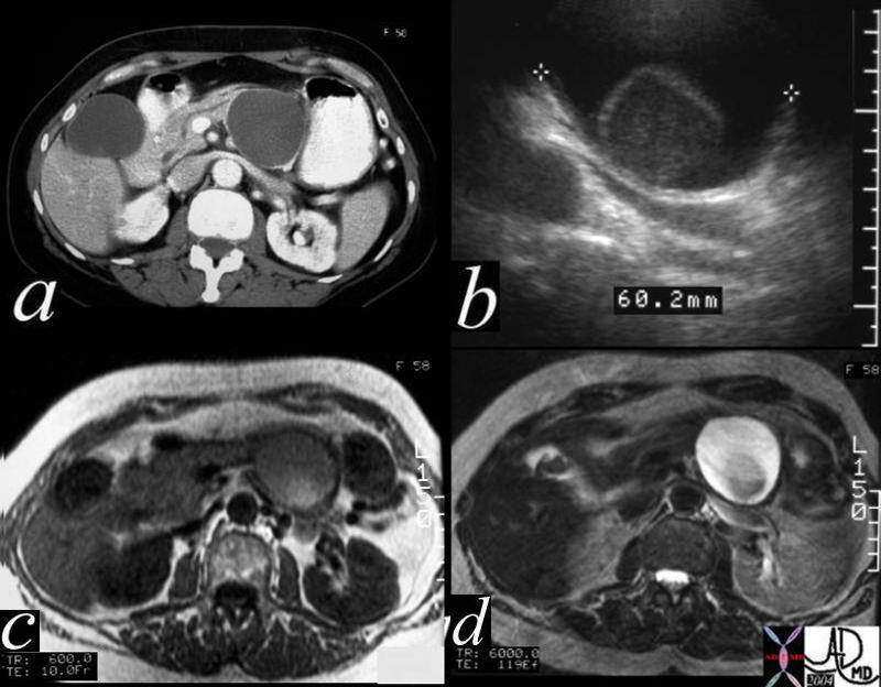
 This combination studies includes CTscans, MRIs, US scans, and an ERCP. They suggest a subtle mass in the head of the pancreas with early bile duct dilatation and more obvious pancreatic duct involvement. This was a case of adenocarcinoma of the head of the pancreas. 41293a16c Courtesy Ashley Davidoff MD
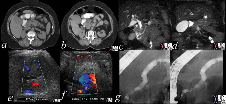
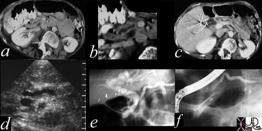 This combination series shows a dilated pancreatic duct in the head of the pancreas (a,b) as well as in the tail (c) The CBD is mildly dilated on US (d) The ERCP (e) shows that both ducts are dilated. At first glance a double duct sign seems likely since both ducts are dilated, but for different reasons. The main pancreatic duct drains into the accessory duct of Santorini, while the CBD drains in usual fashion into the ampulla. The smaller duct of Wirsung is seen mostly disconnected from the ductal system at large. This is a case of pancreas divisum. No pancreatic cancer was found on further workup 40617c01 Courtesy Ashley Davidoff MD
Using CT scan for Fat
DISEASES ON CT
Using CT scan for Acute Pancreatitis
Using CT scan for Carcinoma
This is a classical example of a double duct sign. The dilated pancreatic duct and bile duct almost point to the disease process in this image. A solid mass was identified in the head of the pancreas consistent with adenocarcinoma. 40837 Courtesy Ashley Davidoff MD
The combination of images below reflects the changes of the above case in at different levels and magnification.
This is a classical example of a “double duct” sign. The dilated pancreatic duct and bile duct in a, almost point to the disease process in this image. The mass was as predicted to be in the head of the pancreas. In c and d, a relatively small hypodense mass was identified whose scirrhous nature, and critical location caused early obstructive jaundice. 40840c01 Courtesy Ashley Davidoff MD
This is a series of images from the case history of a 57 year old female who presented with jaundice. The US image (a)
shows a dilated bile duct and dilated pancreatic duct with no large mass between them.
The CT shows a less impressive, but prominent pancreatic duct. The ERCP suggests a “double duct” sign.
The pathology confirmed an ampullary carcinoma. 04655c05 Courtesy Ashley Davidoff MD
This CT study is a variation of the double duct sign. The biliary dilatrelieved by a stent while the pancreatic duct dilatation with secondary atrophy of the pancreas is quite apparent. This is a patient with pancreatic carcinoma.16312
Courtesy Ashley Davidoff MD
This combination studies includes CTscans, MRIs, US scans, and an ERCP. They suggest a subtle mass in the head of the pancreas with early bile duct dilatation and more obvious pancreatic duct involvement. This was a case of adenocarcinoma of the head of the pancreas. 41293a16c Courtesy Ashley Davidoff MD
Using CT scan for Cysts
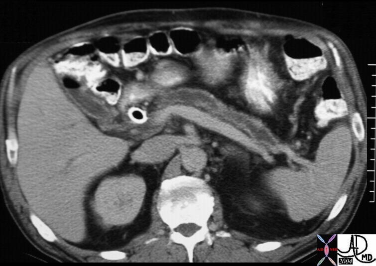
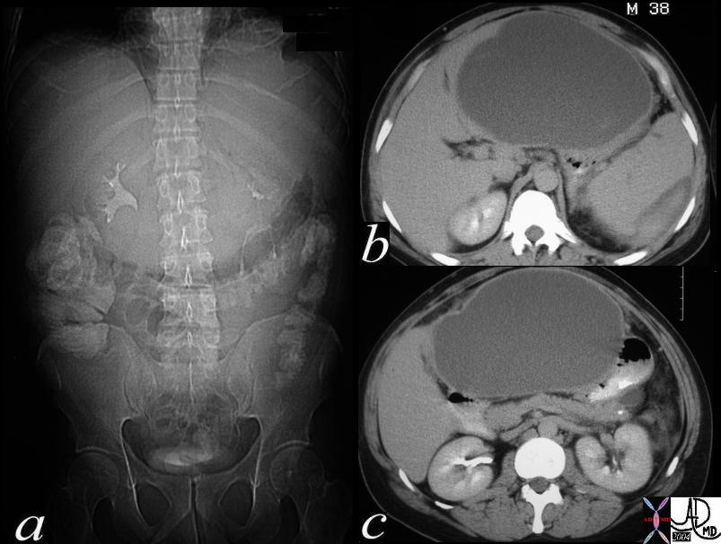 This CTscan is of a 38 year old male with abdominal distension. The scout study shows inferior displacement of the transverse colon by a large epigastric mass. A large cystic structure impinging on the medial left lobe of the liver and the medial aspect of the stomach suggest a pseudocyst arising in the lesser sac from the pancreas and a diagnosis of a pancreatic pseudocyst secondary to acute alcoholic pancreatitis is most likely. The high density material around the spleen in b represents a subcapsular splenic hematoma. 40283c Courtesy Ashley Davidoff MD
This CTscan is through the tail of the pancreas shows what appears to be a simple cyst (a), but which proves to have a mural nodule on ultrasound (b). On the T1 weighted image (c) the nodule shows high intensity suggesting increased protein or acute blood, and the T2 (d) reveals the cystic nature of the matrix. At pathology the lesion was a benign serous cystadenoma. The high intensity on T1 was due to the high protein in the mucin. 40487c01 Courtesy Ashley Davidoff MD
Using CT scan for Pancreas Divisum
This combination series shows a dilated pancreatic duct in the head of the pancreas (a,b) as well as in the tail (c) The CBD is mildly dilated on US (d) The ERCP (e) shows that both ducts are dilated. At first glance a double duct sign seems likely since both ducts are dilated, but for different reasons. The main pancreatic duct drains into the accessory duct of Santorini, while the CBD drains in usual fashion into the ampulla. The smaller duct of Wirsung is seen mostly disconnected from the ductal system at large. This is a case of pancreas divisum. No pancreatic cancer was found on further workup 40617c01 Courtesy Ashley Davidoff MD
Using CT scan for Serous Cystadenoma
Using CT scan for Mucinous Cystadenoma
Using CT scan for Annular Pancreas
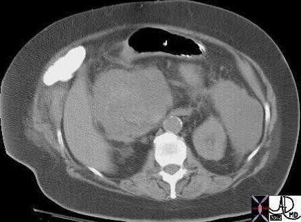 This CT scan through the abdomen shows an accumulation in both paracolic gutters in a patient with acute pancreatitis. The study is performed without contrast and so it gives us the opportunity to observe a hint of increased density in the right sided collection. Narrow windows would be helpful in the confirmation of this finding. Optimal definition of the structures is otherwise difficult without the contrast. A second phlegmonous accumulation is seen in the left side of the retroperitoneum. This is a case of hemorrhagic pancreatitis. 16850 Courtesy Ashley Davidoff MD
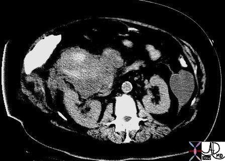 This CT scan through the abdomen shows narrow windows of the accumulated fluid in the paracolic gutters in a patient with acute pancreatitis. The study is performed without contrast and so it gives us the opportunity to observe the increased density in the right sided collection. This is a case of hemorrhagic pancreatitis. 16851b03 Courtesy Ashley Davidoff MD