
The Common Vein Copyright 2008
Introduction
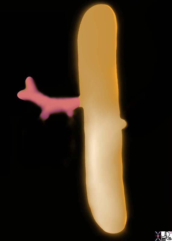
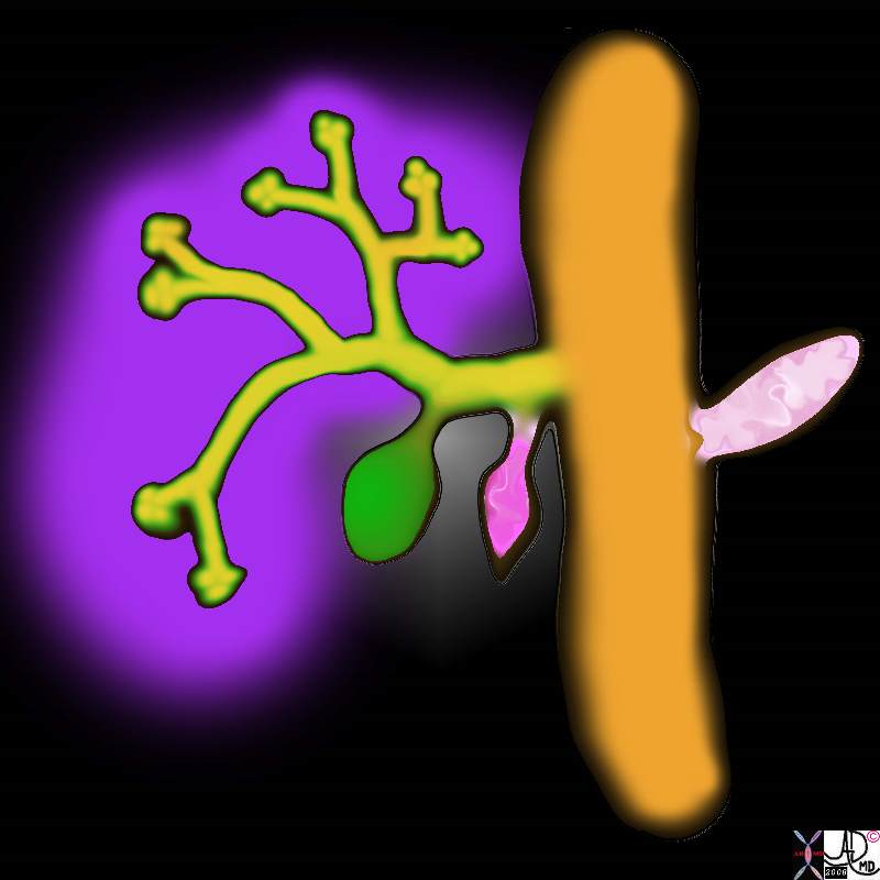
In the 4th week of gestation, the primitive endoderm gives rise to a foregut diverticulum called the hepatic diveticulum at the junction of the of the foregut and midgut. The hepatic diverticulum is the precursor for the liver bile ducts and gallbladder. The hepatic diverticulum gives rise to a pars hepatica and pars cystica. The pars hepatica will evolve into the liver and bile ducts while the pars cystica will develop into the gallbladder and cystic duct. The endoderm bud is initially a solid cord but by week 8 with growth and resorbtion the tubular components are formed
|
|
| In the 4th week of gestation, the primitive endoderm gives rise to a foregut diverticulum called the hepatic diveticulum at the junction of the of the foregut and midgut. The hepatic diverticulum is the precursor for the liver bile ducts and gallbladder. hepatic diverticulum pars hepatica and pars cystica.
liver bile ducts pars cystica gallbladder and cystic duct. endodermal bud solid cord growth and resorbtion gallbladder embryology normal Davidoff art copyright 2008 82219a01b2.8s 82219a02a1.8s |
 Hepatic Diverticulum
|
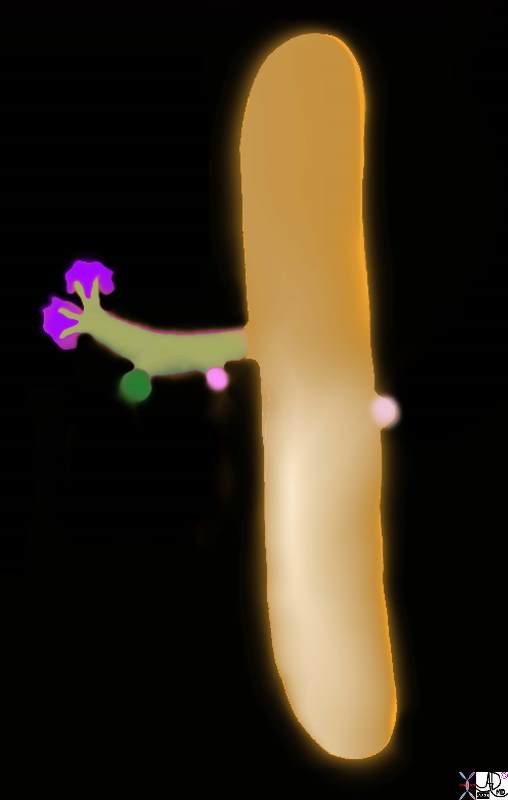
| In the 4th week of gestation, the primitive endoderm gives rise to a foregut diverticulum called the hepatic diveticulum at the junction of the of the foregut and midgut. The hepatic diverticulum is the precursor for the liver bile ducts and gallbladder. hepatic diverticulum pars hepatica and pars cystica.
liver bile ducts pars cystica gallbladder and cystic duct. endodermal bud solid cord growth and resorbtion gallbladder embryology normal Davidoff art copyright 2008 82219a02b1.8s 82219a02b3.8s |
 Hepatic Diverticulum
Hepatic Diverticulum |
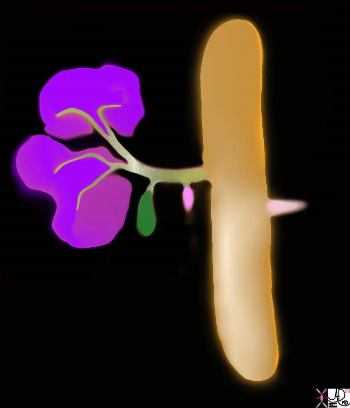
| In the 4th week of gestation, the primitive endoderm gives rise to a foregut diverticulum called the hepatic diveticulum at the junction of the of the foregut and midgut. The hepatic diverticulum is the precursor for the liver bile ducts and gallbladder. hepatic diverticulum pars hepatica and pars cystica.
liver bile ducts pars cystica gallbladder and cystic duct. endodermal bud solid cord growth and resorbtion gallbladder embryology normal Davidoff art copyright 2008 82219b01.8s |
|
|
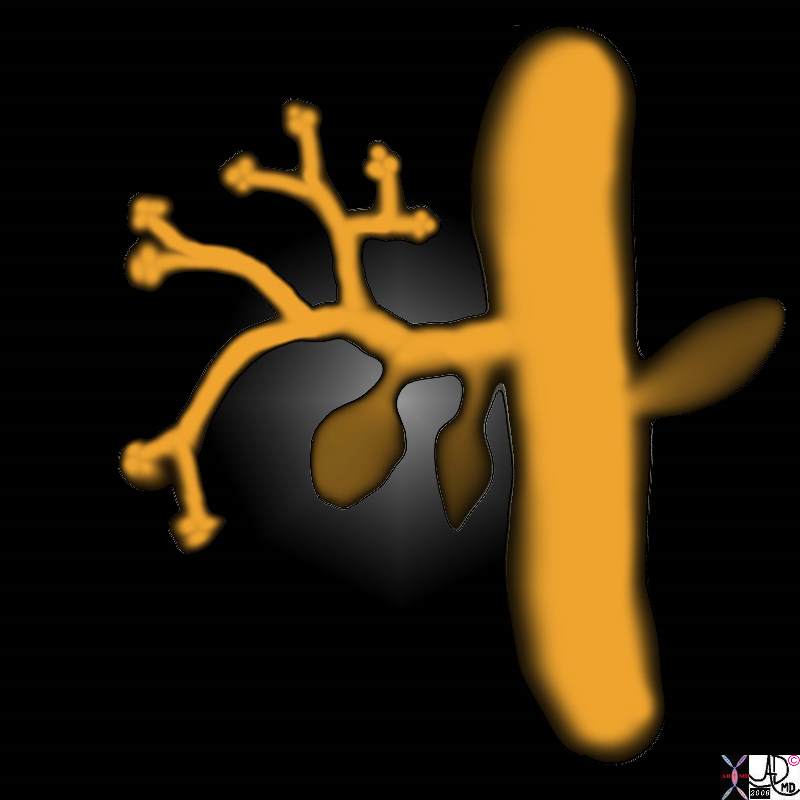
| In the 4th week of gestation, the primitive endoderm gives rise to a foregut diverticulum called the hepatic diveticulum at the junction of the of the foregut and midgut. The hepatic diverticulum is the precursor for the liver bile ducts and gallbladder. hepatic diverticulum pars hepatica and pars cystica.
liver bile ducts pars cystica gallbladder and cystic duct. endodermal bud solid cord growth and resorbtion gallbladder embryology normal Davidoff art copyright 2008 82219b08.8s |
 Hepatic Diverticulum
Dual Origins
Embryologically, the pancreas forms from a ventral anlage that becomes the inferior pancreatic head and uncinate process and from a dorsal anlage that becomes the superior pancreatic head and the body and tail of the pancreas. The fusion of ventrql and dorsal portions occurs at about 6-8 weeks of gestation.
The pancreas arises from two separate endodermal outpouchings from the distal portion of the foregut. These outpouchings originate from opposite sides of the developing duodenum with the ventral portion forming the anlage of the liver, biliary tree, and inferior aspect of the head and uncinate process, while the dorsal anlage grows into the dorsal mesentery to become the tail, body neck and superior portion of the head of the pancreas.
The ventral anlage, in concert with the biliary duct, has to rotate in clockwise fashion around the duodenum in order to enable fusion with the developing dorsal anlage. The fusion occurs between six and eight weeks of pregnancy. While the fusion of parenchyma seems to be seamless, the fusion of the respective ductal systems is more complex and results in a variety of morphogical complexes.
The usual result of the fusion is a minor duct that drains the superior aspect of the head and a major duct that drains threst of the pancreas.
The portion of the dorsal duct that had formerly drained the head of the pancreas becomes the minor duct of Santorini and it drains the superior portion of the head and enters the duodenum about 1-2cms cranial to the papilla. The major pancreatic duct of Wirsung is composed of the the dorsal duct subtending the tail and the body and the ventral duct draining the inferior portion of the head and uncinate. The relationship of the ventral potion of the pancreatic duct and the the common bile duct remains constant and the junction forms the ampulla which terminates in the papilla within the mid portion of the descending duodenum.
Duality of the Glands
The side branches from the main duct derive from buds of the main ducts and give rise to blind ending to the tubules, which evolve into the acini. The ductal epithlium is multipotential and gives rise to both the acinar cells and islet cells. The islets lose their connection with the ductal system as they evolve and connect with the vascular system which eventually supports their needs as an endocrine organ. The multipotental ability to differentiate into acinar or islet morphology is apparrently maintained into adult life.
There is a normal variation of the shape of the head based on the dominance and relative contibutions of the ventral and dorsal portions of the pancreas.
 The ventral remnant has a variable shape as it relates to the duodenum. It is mostly an inverted “c”
and occupies about 1/3 to 1/4 of the circumference. (a-f). Sometimes it partially or totally surrounds the
duodenum resulting in the entity of annular pancreas (h) 41505c02 Courtesy Ashley Davidoff MD
Annular pancreas
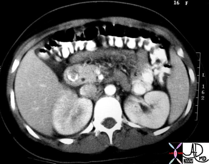 In this cross sectional image of the pancreas the ventral portion of the pancreas totally surrounds the duodenum
but does not cause obstruction. This is a case of annular pancreas. 30906a04 Courtesy Ashley Davidoff MD
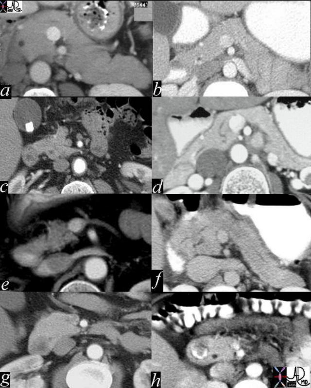
 This is a CT scan through the pancreas where there is only a partial divisum anomaly. Images a and c are two different
levels showing almost 70% encirclement of the pancreas (pink) around the duodenum (d orange).
Courtesy Ashley Davidoff MD 18090c06
Pancreas Divisum
Pancreas divisum, represents the failure of fusion of the two ductal systems so that the dorsal duct of the tail body and superior portion of the head becomes the main system draining into the minor papilla while the ventral system drains only the head and uncinate process but maintains its relationship with the CBD. This is not an uncommon anomaly occurring in about 1 in 12 people and most often has no clinical consequences. It can however lead o pancreatitis presumably due to reflux of duodenal contents into the pancreas. The dorsal duct usually loses its connection and becomes the accessory duct of Santorini.
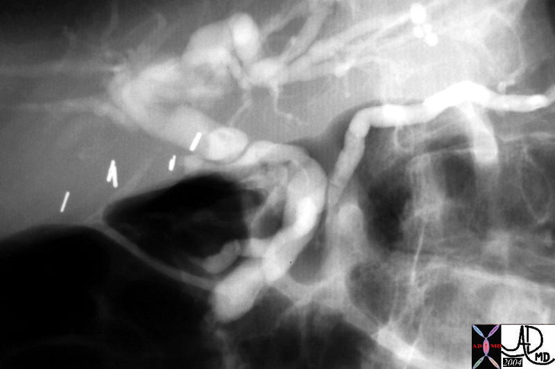 This a case of pancreas divisum as seen by ERCP. The MPD is continuous with the APD of Santorini as it enters the
duodenum 1-2cms cranial to the papilla and the entrance of the CBD. 40615 Courtesy Ashley Davidoff MD
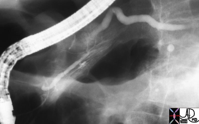 This is the injection of the MPD of the case above showing a small ventral duct of Wirsung. The case represents pancreas divisum as seen by ERCP. The main duct was found opening separately within the pailla near the opening of the CBD. 40617 Courtesy Ashley Davidoff MD
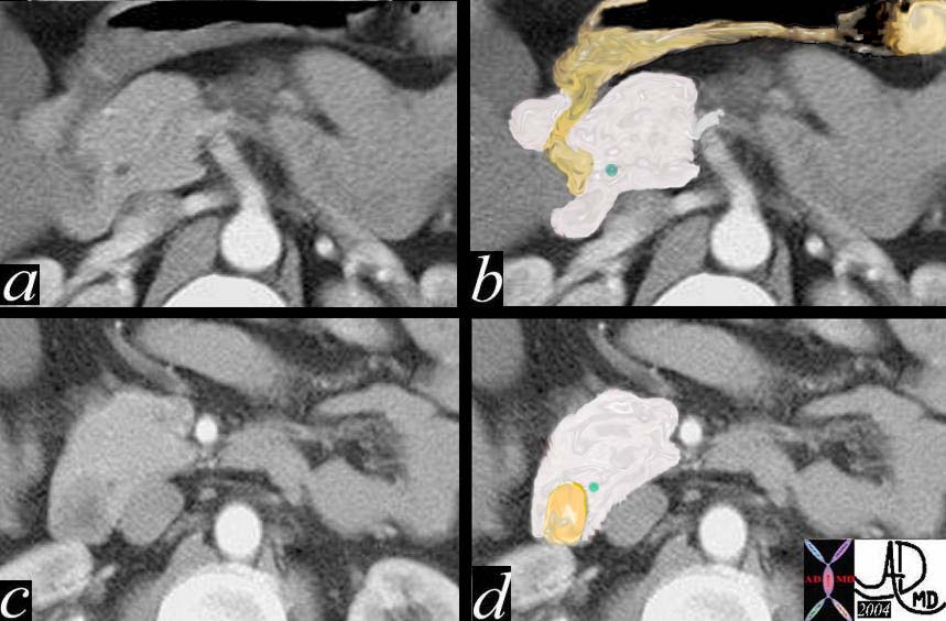
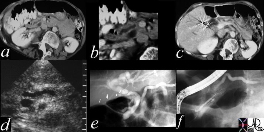 This combination study of pancreas divisum consists of a CTscan with the MPD by passing the CBD (a) magnified in b, with a slightly dilated appearance in the body of the pancreas in c. The US equivalence of this finding is noted in d, while e shows the MPD and ventral duct exiting cranila to the entrance of the CBD. The last injection of the ERCP is the injection of the MPD showing a small ventral duct of Wirsung. The findings are pathognomonic of pancreas divisum. Courtesy Ashley Davidoff MD 40617 c01