| The Common Vein Copyright 2007
From the radiological point of view, the pancreas is less challenging than for the clinician. CTscan offers the single best window to the pancreas, and endoscopic ultrasound the single best focused view of the pancreas. The pancreas was known as the hermit of the abdomen, and in many ways it is till a truism since it hides itself behind the stomach and in front of the spine and is protected on either side by antrum duodenum small bowel and large bowel. However with the advent of cross sectional technology, the pancreas has been exposed to the imaging eye .
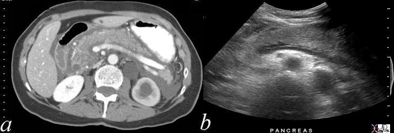 Acute Pancreatitis – CT and Ultrasound Acute Pancreatitis – CT and Ultrasound |
| 70369c02 pancreas acute pancreatitis enlarged fluid interstitial fluid third spacing 3rd spacing CTscan USscan Davidoff MD 70369c01 |
Each technique in imaging has something to offer. The plain film of the abdomen is perfect for making the diagnosis of chronic pancreatitis when the pancreas is heavily calcified but usually the diagnosis at this stage is well established clinically. The signs of acute pancreatitis which include sentinel loop characterized by a focal ileus or colon cut-off representing focal obstruction of the splenic flexures are both very helpful in the patient presenting with abdominal pain
US is a good overview examination for the patient with abdominal pain and for the characterization of masses, particularly cystic masses, but the central location of the pancreas behind the stomach small bowel and sometimes behind colon makes the study inconsistent since gas inhibits and reflects the interrogating ultrasound waves . Endoscopic ultrasound is great for the identification and biopsy of small tumors, and can also be used for the drainage of cysts and the pancreatic duct. It suffers with its focused view to appreciate the big picture.
CTscan offers the single best study of the pancreas for its full and consistent view of the pancreas and surrounding structures, while MRI is not far behind providing a better view of the pancreatic and bile ducts.
ERCP has the unique advantage of enabling the gastroenterologist the ability to intervene and treat many of the obstructive conditions that may affect the biliary and pancreatic tree.
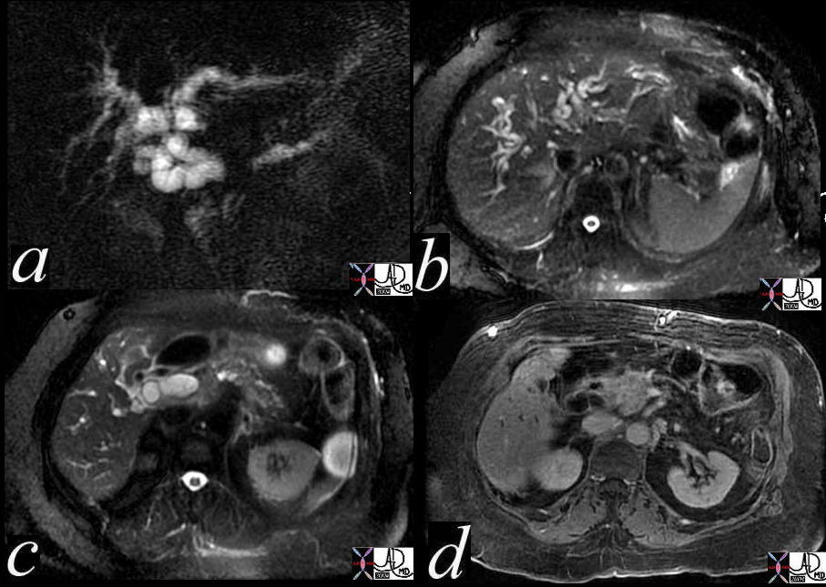 Double Duct Sign Double Duct Sign |
| In the MRCP (a) the double duct sign is well demonstrated with a dilated intrahepatic bile duct, and a dialted pancreatic duct. The axila images (b,c) show dilated intrahepatci ducts, while the gadolinium enhanced image (d) shows a relatively vascular mass in the body and neck of the pancreas. 41371c Courtesy of Ashley Davidoff MD |
The octreotide scan is a radioisotope study that is sensitive to neuroendocrine tumors that have somatostatin receptors. In the octreotide scan an antibody to this receptor is labeled with a radioisotope. Tumors that are rich with these receptors will have many of the antibodies bind to them. Many neuroendocrine tumors such as a gastrinoma or an insulinoma of the pancreas have a high concentration of the somatostatin receptors compared to normal tissue. The radioisotope will be selectively attracted to such a tumor and present as a “hot spot”. When positive, this study is extremely sensitive and specific.
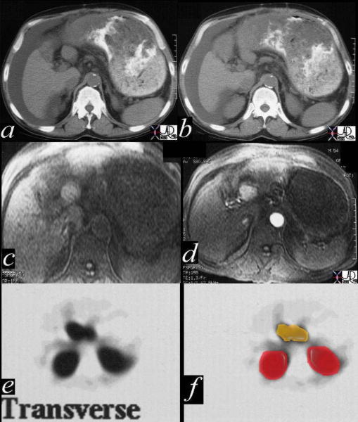 This combination series is from a patient who was presenting for liver transplant who demonstrated an isodense mass noted on CT (a,b) that was thought to represent a regenerating nodule. He had an unexplained gastropathy or gastric oulet syndrome, characterised by a stomach that was always filled with complex secretion. An MRI (c,d) showed an non-specific enhancing mass in the porta hepatis. The octreotide showed a hot spot in the same location (d,e) with the gastrinoma noted in yellow, while the enhancing kidneys noted with red overlay. Courtesy Ashley Davidoff MD 19644ec This combination series is from a patient who was presenting for liver transplant who demonstrated an isodense mass noted on CT (a,b) that was thought to represent a regenerating nodule. He had an unexplained gastropathy or gastric oulet syndrome, characterised by a stomach that was always filled with complex secretion. An MRI (c,d) showed an non-specific enhancing mass in the porta hepatis. The octreotide showed a hot spot in the same location (d,e) with the gastrinoma noted in yellow, while the enhancing kidneys noted with red overlay. Courtesy Ashley Davidoff MD 19644ec
PET scanning is now evolving as a study with high sensitivity and specificty for the diagnosis of carcinoma. Time will tell whether it is helpful in making an outcome difference in carcinoma |
