
Author Ashley Davidoff MD
Collaborators Charles Allison MD Adam Asarch MSII David Lee MD Scott Tsai MD Sam Yam PhD.
Approach
Since most are related to alcoholic pancreatitis, the challenge is to identify a cause when there is no history of alcohol abuse.
Differential Diagnosis
chronic alcoholic pancreatitis (85-90%)
Situated in the ducts –
punctate or diffuse in ducts
hereditary pancreatitis
hemorrhage/hematoma
tumors
serous cystadenoma
benign, female, tiny cysts, (but may be up to 2cms) large tumor, may be head body or tail, central stellate scar, +/- calcification
mucinous cystadenoma/ cystadenocarcinoma
benign, premalignant, or malignant, female++, large (mean 12cms) thick septations, 85% tail and body, scattered dystrophic calcifcation
cystic fibrosis
hemochromatosis
idiopathic
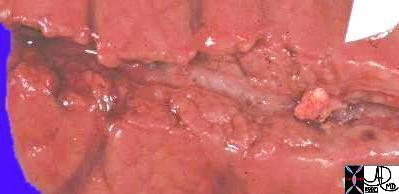
This grosspathology specimen shows a calcified stone within the pancreatic duct. This is almost pathognomonic fro alcoholic pancreatitis. 15303c Courtesy Barbara Banner MD
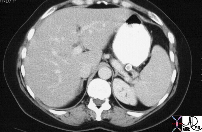
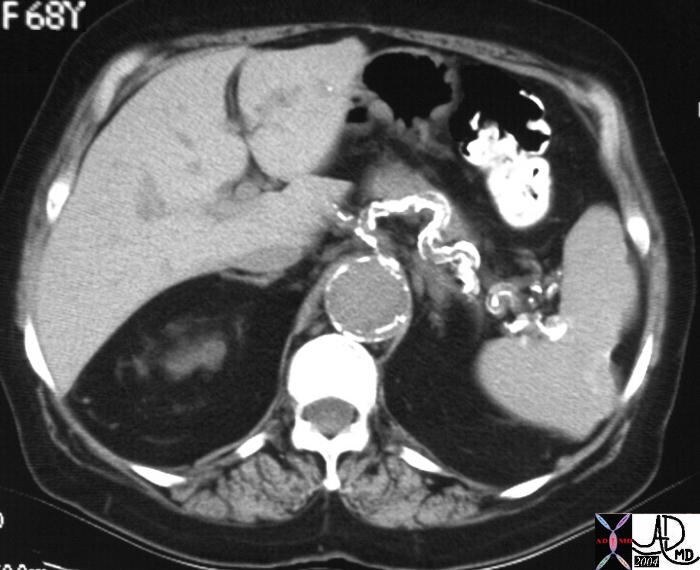 The splenic artery running above the pancreas is heavily calcified in the CT scan of this 68 year female. The splenic artery has a serpiginous shape and is one of the first vessels to calcify in the abdomen. It is therefore not uncommon to have calcifications of the artery masquerade as calcifications of the pancreas. The clue to the arterial origin of of the calcification is usually the linear nature of the calcification. Associated finding in this case is a scar in the spleen which is associated with calcification within and on the surface of the spleen. 22014b. Courtesy Ashley Davidoff MD
 Small splenic artery aneurysms are not uncommon in the elderly and since the artery is so intimately related to the pancreas, one may mistake the aneurysm for a calcified mass of the pancreas. The aneurysm is usually classically located within a few cms of the hilum of the spleen, is mostly almost always round with a thick rind of circumferential coarse calcification. 20657 Courtesy Ashley Davidoff MD
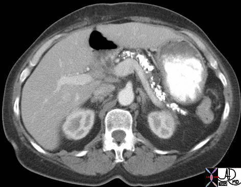
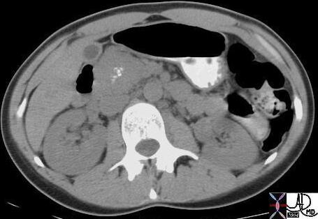
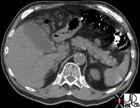 The CT through the abdomen shows punctate calcifications scattered in the periphery of the pancreas and most likely in the secondary ducts of the pancreas. The findings are most consistent with chronic alcoholic pancreatitis. In addition there is a tiny calcification in the infundibulum of the gallbladder associated with mild induration of the gallbladder. These findings are consistent with acute cholecystitis. The calcifications in the periphery of the spleen are unusual and are of unknown significance. 40761 Courtesy Ashley Davidoff MD
 This 58 year old female presents with recurrent acute episodes of severe abdominal pain. This CTscan shows heavy calcification of the body and tail with a segmenin the middle of a dilated pancreatic duct. The findings are consistent with chronic alcoholic pancreatitis. 41241 Courtesy Ashley
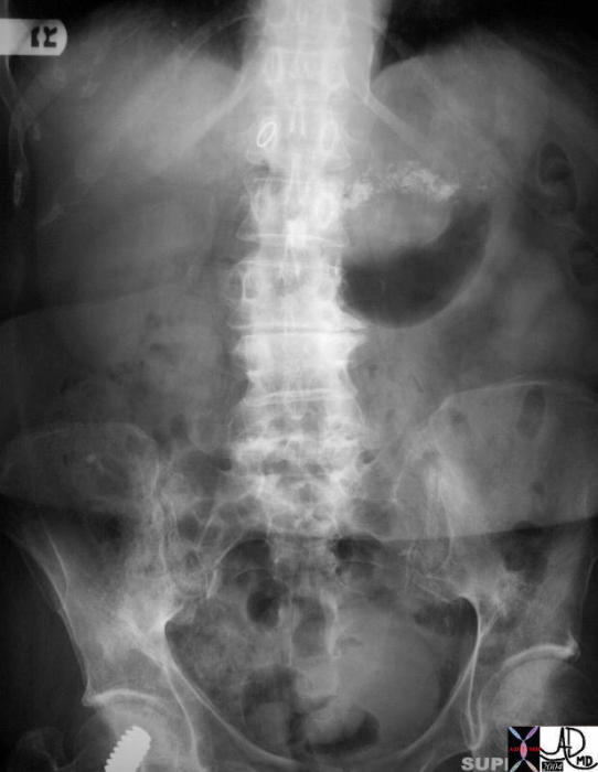 This plain film of the abdomen shows the tubular shaped calcification of the pancreas which lies in the transpyloric plane. Incidental finding is a compression screw in the right hip. The most common and likely diagnosis is chronic pancreatitis 32913 Courtesy Ashley Davidoff MD
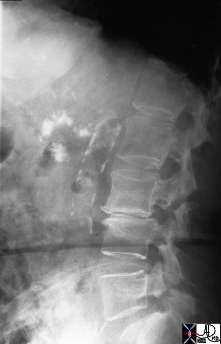
The patient above also had a lateral examination which shows the relative posterior and high position of the tail, and the inferior and anterior position of the uncinate process. Incidental findings include the calcified atherosclerotic abdominal aorta and the degenerative changes at L3-L4. 32916 Courtesy Ashley Davidoff MD
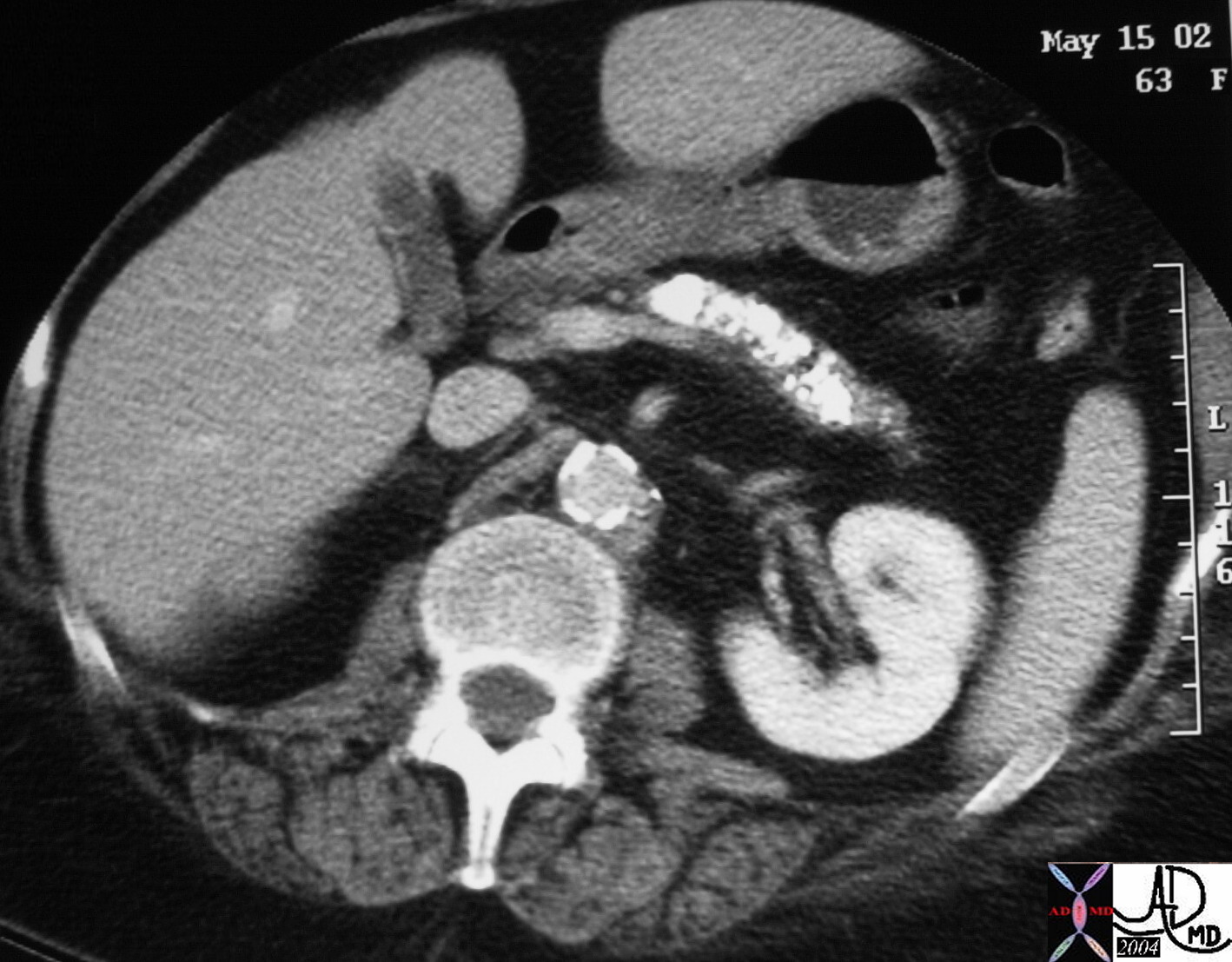
 The CT scan of the abdomen of a 63year old female shows dense calcification of a small pancreas, characteristic of chronic alcoholic pancreatitis. Associated finding includes a low density filling defect in the gallbladder that probably represents a cholesterol stone 32920 Courtesy Ashley Davidoff MD
 The CTscan through the head of the pancreas shows a punctate calcification in the region of the head of the pancreas in close contact with the cystic appearing CBD. Associated findings include induration around the head of the pancreas and thickening of the left sided Gerota’s fascia. These findings are consistent with a diagnosis of gallstone pancreatitis. 16225b03 Courtesy Ashley Davidoff MD
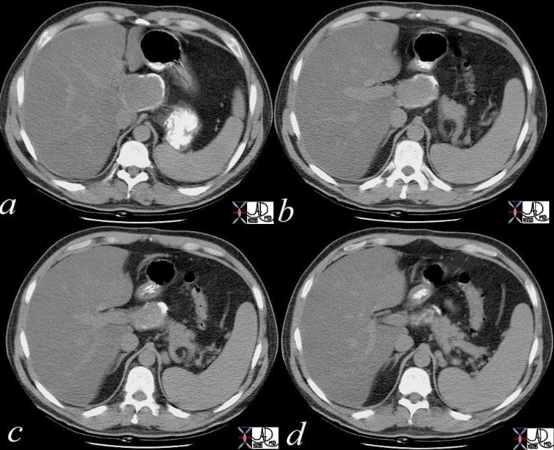
 The CTscan through the body of the pancreas shows a dilated pancreatic duct with two punctate radio-opacities, associated with high lying small bowel in the gallbladder fossa. This patient had surgical removal of the head of the pancreas and the small bowel was brought up to drain the biliary system and pancreatic system. The punctate densities probably represent sutures. 40319c Courtesy Ashley Davidoff MD
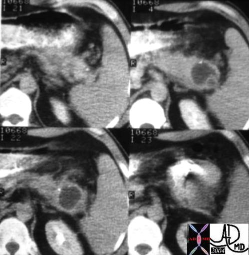 The CTscan through the body of the pancreas is of a 74 year old female with abdominal discomfort. She has no history of alcohol abuse and a normal US of the gallbladder. The study shows a complex, bilocular, cystic lesion in the tail of the pancreas with an enhancing rind, and two focal peripheral punctate calcifications. A diagnosis of a cystadenoma is thought to be most likely. 04811 Courtesy Ashley Davidoff MD
 The CTscan through the head of the pancreas is of a patient with known von Hippel-Lindau syndrome. It shows a solid mass with central dystrophic calcification in the head. In this setting the diagnosis is most likely a serous cystadenoma. 18378 Courtesy Ashley Davidoff MD The CTscan through the head of the pancreas is of a patient with known von Hippel-Lindau syndrome. It shows a solid mass with central dystrophic calcification in the head. In this setting the diagnosis is most likely a serous cystadenoma. 18378 Courtesy Ashley Davidoff MD
|
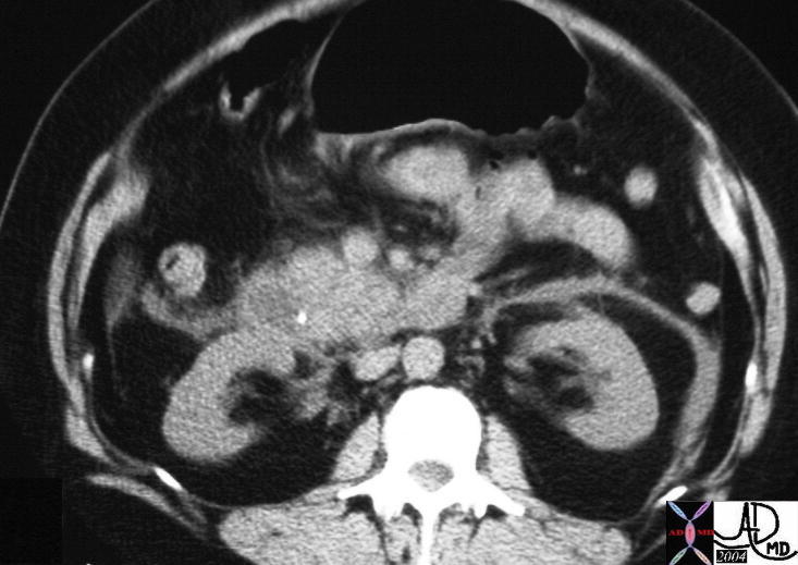
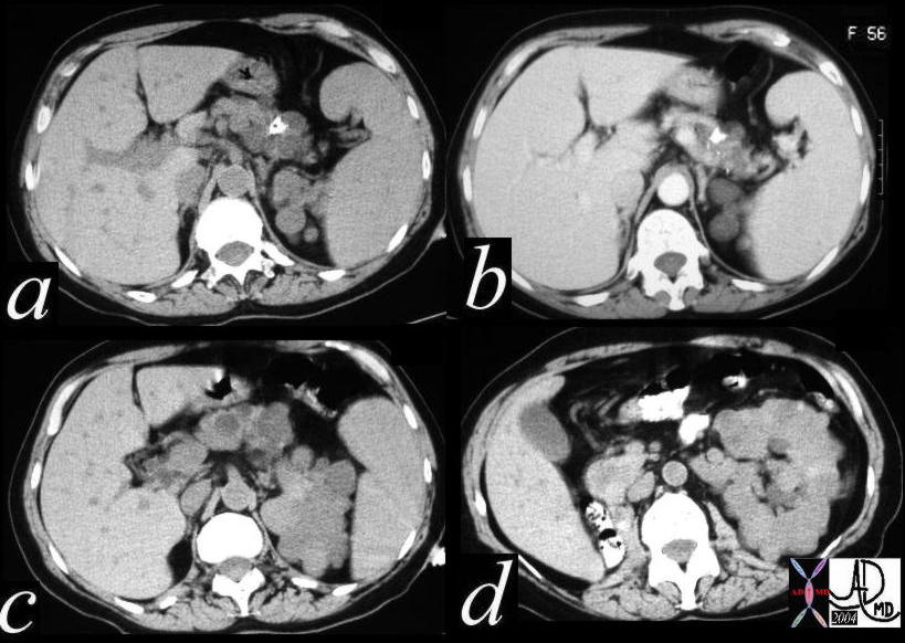 This CT scan through the pancreas shows a complex cystic mass with central calcification in the tail of the pancreas associated with complex masses in the left kidney and surgically absent right kidney. These findings are consistent with a diagnosis of von Hippel-Lindau syndrome with a variety of cystic and solid, benign and malignant tumors of the pancreas and kidney. 40463c Courtesy Ashley Davidoff MD
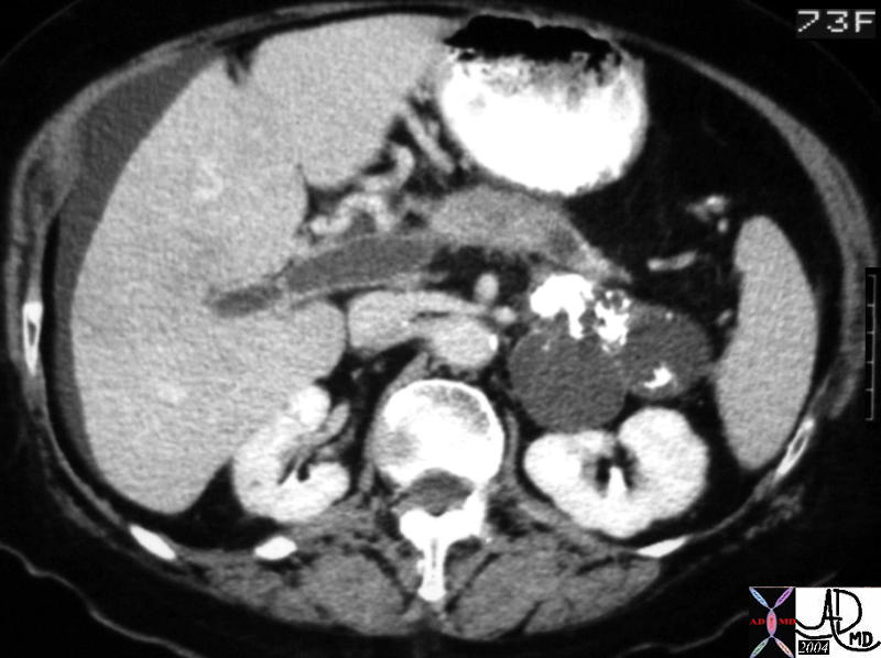 This CT scan through the pancreas shows a complex cystic mass with dense peripheral dystrophic calcification in the tail of the pancreas. Associated findings include portal vein thrombosis (PVT) and ascites. These findings are consistent with a cystadenoma, and a malignant transformation of a mucinous neoplasm has to be considered in view of the PVT. An expanded version of this case appears below. 40712 Courtesy Ashley Davidoff MD
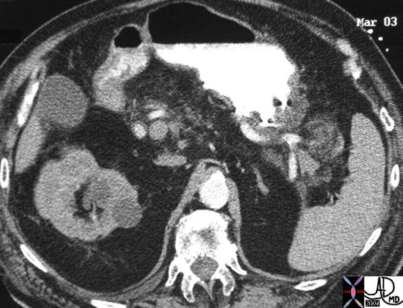
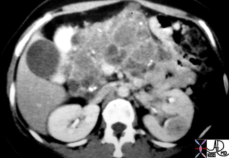 This CT scan through the pancreas shows a series of complex cystic and solid masses mass with punctate calcifications throughout the whole pancreas associated with a solid mass in the left kidney. These findings are consistent with a diagnosis of von Hippel-Lindau syndrome with a variety of cystic and solid, benign and malignant tumors of the pancreas 40531 Courtesy Ashley Davidoff MD
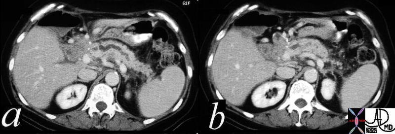
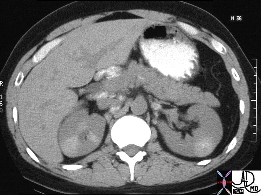 This CT scan through the pancreas shows a series of calcified masses surrounding the head of the pancreas but also in the porta hepatis, portocaval space, and paracaval position. These findings represent lightly calcified lymph nodes. Associated findings include unusual wedge shaped calcifications in the left kidney, and mass like calcification in the right. This is a patient with sarcoidosis. The lymph node pattern in the foregut is not unusual for sarcoidosis. Kidney involvement with granulomatous disease is not uncommon, but the pattern of calcification in the kidney is quite unusual. 26477b02 Courtesy Ashley Davidoff MD
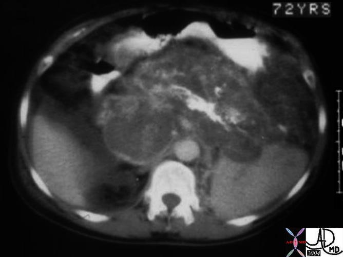 This CTscan of a 72 year old man through the body of an enlarged pancreas shows a central almost linear calcification with surrounding low density, representing the bulk of the parenchyma. Strands of soft tissue and blood vessels are scattered in the rest of the gland. At surgery this proved to be a giant hemangioma of the pancreas. This is not your usual case!! 04894c001 Courtesy Ashley Davidoff MD