Celsus in the first century AD described a condition likely to have been diabetes, Noting “excessive pouring out of urine” and causing “emaciation and danger.” Galen emphasized thirst, while sweet urine was described by the Hindus in the Brahman period in 500AD and later by the Englishman, Thomas Willis in the late 1600’s. Another Englishman, Matthew Dobson noted the sweetness of both the urine and the blood about 100 years after Willis. It is very likely based on description that these physicians went so far in the clinical exam as to taste the urine of their patients! Talk about dedication to diagnosis, patient and research! The clinical clues of polydydypsia, polyphagia and polyuria remain the hallmarks of the clinical diagnosis of diabetes. The presence of jaundice and an enlarged gallbladder is reminiscent of the Courvoisier law, which states that that ‘if in the presence of jaundice the gallbladder is palpable, then the jaundice is unlikely to be due to a stone.’ The likely cause of this entity is therefore cancer in the head of the pancreas. Clay colored stools and dark urine would go along with the diagnosis of obstructive jaundice. These tools are clues but occur relatively late in the disease.
| Historical Aspects
The Common Vein Copyright 2007
Introduction
In this section we explore the evolution of pancreatic knowledge with particular emphasis on structure, from the time of antiquity to the current day. 300-400 BC on the Asiatic side of the Bosporus river. Herophilus was one of the founders of the ancient schools of Medicine in Alexandria, Egypt. He may have been the first to have performed dissections of human bodies before public audiences, and thus is considered the father of scientific anatomy. He accurately described the eye, brain, liver, and pancreas, though the name “pancreas” only seemed to have evolved later. The pancreas appears in Aristotle’s Historia Animalium, in which he refers to “another to the so-called pancreas” suggesting that not only was the organ known to Aristotle, (384-322 BC) but the name was already established. In the English translation of chapter 4 by Wentworth Thompson of Historia Animalium the word pancreas seems to be mentioned in context …”Again, other veins branch off from the big vein; one to the omentum, and another to the pancreas, from which vein run a number of veins through the mesentery. All these veins coalesce in a single large vein, along the entire gut and stomach to the oesophagus; about these parts there is a great ramification of branch veins.” Historically however Ruphos an anatomist who lived in the first or second century AD has been given the credit for assigning the name to the pancreas. He was a surgeon of Ephesus in Asia Minor which today is the Asian portion of Turkey. The name “pancreas” means “all meat” or “all flesh” in Greek. 100-200 AD 300 AD 1200-1300 1400 AD
1500 AD
Andreas Vesalius (1515-1564) was born in Brussels, and educated in Paris and Padua as an anatomist and surgeon. Many consider Vesalius the true father of anatomy. He emphasised that the study of anatomy should come from direct observation from the human body and not learned from animals (which had been the habit of Galen) nor only from books (which had been the habit of others for so many years) . Vesalius’s brought a number of radical changes to the study of anatomy, but despite his open minded approach, and his disagreement with Galen on at least 200 other anatomic issues, he continued to follow Galenic theory that the pancreas functioned as a support structure for the stomach and mesenteric vessels. There was therefore no significant advancement on pancreatic knowledge in his time. His illustrated book De Hummani Corporis Fabrica was published in 1543. In plate 54 he noted that the “glandular” bodies arising in it” inferring that the pancreas arose in the the omentum and transverse mesocolon. The pancreas was regarded as several glands probably beacause it was broken up by the dissection of the vessels that course in and around it. In plate 56 there is a diagram of the greater omentum and transverse mesocolon where there is an elevation thought to be a representation of the pancreas and labelled with an “L”. 1600
1632 The Anatomy Lecture of Dr. Nicolaas Tulp (1593-1674) by Rembrandt Harmenszoon van Rijn (1606-1669) Dr Tulp was one of the first to identify the anatomical changes of pancreatitis in a post mortem. In this particular painting he is dissecting the upper limb of a criminal who had been hanged for his crime. Aris Kindt had been hanged for armed robbery. Immediately after the execution, his body was taken to the Anatomy Theatre of the Guild of Surgeons. 54466 code historical reference Thre is a great explanation of this image at http://www.ohsu.edu/library/images/hom/rutkow122.jpg In 1642, a German émigré to Italy and prosector Johann Georg Wirsüng, discovered the pancreatic duct when he was a medical student in Padua, Italy. He questioned it’s nature and function and observed that there was never blood within it. He engraved a drawing of the duct on a copper plate and made seven copies, distributing the plates to other well known anatomists in Europe with the question as to whether it was an artery or a vein. Wirsüng was murdered by a student the year after his discovery. (Howard ) It was rumored that he was murdered by a his jealous mentor Johann Wesling, but Wesling was subsequently acquitted of the crime. Wirsung’s diagram of the duct resides in the University of Gottingen.
At about the same time in England, in the middle of the 17th century, Thomas Wharton an anatomist, was also focused on the glands of the body, culminating in a Latin treatise Adenographia published in 1656. The work covered a description of the glands of the body providing the first thorough account and focus on the presence and importance of the glands. More importantly he distinguished them from the other viscera. He was able to classify the glands into excretory, reductive, or nutrient types. Wharton noted the superficial similarity between the pancreas and the submaxillary salivary glands. He discovered the duct of the submandibular salivary gland, which was subsequently called Whartons duct. He did not take the obvious next step to find the duct of the pancreas. It would have been unlikely that Wharton was consulted by Wirsung about the nature of the bloodless tube of the pancreas since he would undoubtedly have known the answer. Not only had he noted the similarity of the structure of the pancreas with the salivary glands but also had discovered the duct of the submandibular gland. It did not take too much longer to understand the nature of the bloodless duct since de Graaf in 1664 in Holland had isolated the duct in the dog and was gaining early insight into pancreatic function.
Reignier de Graaf (1641-1673) of Holland wrote a treatise in latin which was later translated into English It was called Tractatus Anatomico-Medicus De Succo Pancreatico, – A physical and anatomical treatise on the nature and office of the pancreatic juice. It is one of the, if not the earliest works on pancreatic secretion. It seems that de Graaf and Wharton independantly came to the conclusion that the pancreas was a gland rather than an organ. This conceptual change, was an important step in the biological knowledge of the glands. De Graaf’s first work was originally published in Latin in 1664. He used the hollow quill of a goose feather to cannulate the pancreatic duct of a dog in 1663. He was the first to isolate and collect the secretions of the pancreas and gall bladder. Apparently he deduced that pancreatic juice was acidic based on his observation upon tasting it, that it was “insipid” with “acid-salt” flavor. He also collected the pancreatic juice of a sailor who died suddenly and noted that it was similar in character to the dogs juices. In 1654 Francis Glisson, (1597-1677 ) published the first book devoted exclusively to the anatomy of the liver. Glisson injected coloured liquids enabling him to outline the vessels of the liver. He was thus able to identify and be the first to describe sphincter of the bile duct (1681) which was later rediscovered by Oddi in 1869 and is known today as the sphincter of Oddi. In 1673, the Swiss physician Johann Conrad Brunner, while still a student in Paris, came near to discovering pancreatic diabetes after he noted that an experimental dog became thirsty and polyuric after a pancreatectomy. His book ‘Experimenta nova circa pancreas’ was published in 1683. The term “diabetes mellitus” was coined in 1674 by the Oxford physician Thomas Willis, personal physician to King Charles II. Mellitus is Latin for honey, which is how Willis described the taste of the urine of diabetics (“as if imbued with honey and sugar”). Thomas Willis also described the arteries at the base of the brain. 1700AD Giovanni Santorini (1681-1737) of Italy studied medicine at Bologna, Padua, and Pisa, receiving the doctorate in 1701. One of his teachers was Marcello Malpighi (1628-1694). Santorini described the pancreatic accessory duct in 1724. 1800 AD.
The two diagrams show the book cover of Claude Bernard’s book on expiremetal medicine and an etching by R. de Los Rios, after a painting by Leon Augustin L’hermitte, 1889. reference UTMB.edu In 1847 Claude Bernard began a series of experiments that involved feeding famished rabbits high protein diets. On the basis of these experiments he was able to deduce that the secretions of the pancreas changed th ingested fat into fatty acids and glycerine. He attributed the reactions to an enzyme that was later called ‘pancreatic lipase’ Despite the advancements made in physiology, it was not until 1852, almost 70 years after the introduction of Leeuwenhoek’s microcopes, that a student in Paris named D. Moyse, pioneered the knowledge of pancreatic acinar histology. Rugerio Oddi (1864-1913) was an Italian anatomist and surgeon of Perugia who described the sphincter bearing his name in 1869. He struggled wih addictions throughout his life and was dismissed from his post as head of the Physiology Institute at the University of Genoa because of impropriety with drugs and finacial issues. Glisson had described the sphincter in 1681 but it may have been too early for the field to grasp and understand the context and importance and thus Oddi was given the honor of having his name associated with the sphincter.
Courtesy Ashley Davidoff MD Alberti, Schenck and Tulp had noted post mortem changes in the diseased pancreas in late 16th and early 17th century and after Claude Bernard’s work on the lipolytic function (1847) the pathology of fat necrosis in acute pancreatitis was recognised by W. Balser in 1882, while autodigestion was a process recognised by H. Chiari in 1896.
In 1869, at an inaugral dissertation of his doctorate degree, Paul Langerhans, (1847-1888), presented his work on the islets of the pancreas subsequently known as the “islets of Langerhans” which represent only 2% of the cellular population of the pancreas. His work was conducted at the Berlin Institute of Pathology, under Rudolph Virchow. Langerhans, a physician, pathologist and anatomist, was innovative in the use of special stains which enabled visualisation of structural detail not before appreciated. The special stains allowed him to recognise the dendritic cells in the skin which now bear his name, and subsequently the islets. Identification of the islets opened up the door (or gates) to a huge field of knowledge relating to glucose metabolism and diabetes.
A picture of Paul Langerhans who discovered the islets of Langerhans while working in Berlin under Virchow Image from Historical Medical Personalities Langerhans contracted tuberculosis of the lung which forced him to emigrate to Madeira in Portugal where he continued with unbridled intellectual enthusiasm and he changed the course of his academic direction by exploring marine fauna of fthe Portuguese coast . He eventually died in Madeira of a renal infection at the age of 41 years in 1888. Claude Bernard had laid the foundation of experimental physiology of the pancreas in 1847, and subsequent collaborators in Johann Nepomuk Eberle (1798-1834) of Bavaria, Alexander Danilevsky of St. Petersburg (in 1862), and Willy Kuhne (1837-1900), of Amsterdam advanced our knowledge of the pancreatic digestive enzymes.
In 1889 as the 19th century was closing, Oskar Minkowski, (1858-1931) a German internist and physiologist and Joseph von Mering (1849-1908) a pharmacist, were debating about whether pancreatic enzymes play a role in the digestion of fat. It was Joseph von Mering who suggested the idea of the pancreatectomy and thought it impossible, while Minkowski a German internist and physiologist became surgeon and removed the pancreas. Actually Johann Conrad Brunner in 1673 had already observed that pancretectomy had resulted in polyuria and polydypsia. Carl Gussenbauer (1842–1903), was a surgeon who pioneered pancreatic surgery. In 1882 he performed external drainage for a pancreatic cyst. Gussenbauer initiated the modern era of pancreatic surgery. Würzburg, Germany
Wilhelm Roentgen more than anybody else is responsible for initiating the work that subsequently opened up the in vivo secrets of the structural makeup of the pancreas. Image may be subject to copyright web 1900 AD The connection between cholelithiasis and acute pancreatitis (E.L. Opie and W. St. Halsted 1901) laid the foundation for 20th century research. In 1902, W.B. Bayliss and E.H. Starling, of London England discovered secretin, which stimulates pancreatic enzyme secretion. References of Historical Aspects John M. Howard, M.D. |

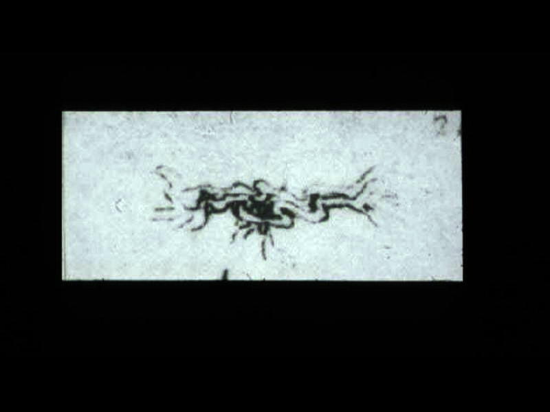 This is a drawing of the “meseraic vessels” – presumably the celiac axis. Da Vinci was able to interview and perform an autopsy on a centenarian. He describes the dessicated and tortuos state of the vessels of this patient accurately depicting the atherosclerotic process. He describes the narrowed lumen and the consequences of poor blood flow.The translation of da Vinci’s text accompanying this one inch image is remarkably insightful and pioneering. (see atherosclerosis hisrorical da Vinci) 13045b
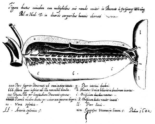 54476 duct of Wirsung code pancreas duct of Wirsung anatomy historical drawing The image depicted is in the University of Gottingen and was photographed by Professor Doctor Heinz Becker, Chief of Surgery. reference – Howard JM, et al., J Am Coll Surg 1998;187201-211. need to obtain permision
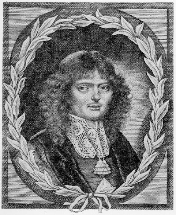 54479a Reinier de Graaf approximately 23 years. The portrait was drawn by his girlfriend, Anna Maria van Schurman around 1664. At the time he was studying in Utrecht, the Netherlands. 54479 web copyright unknown ref
 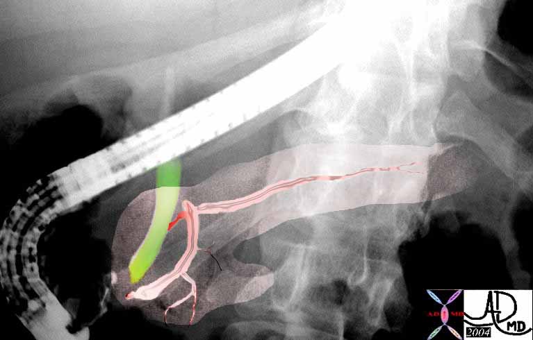 This ERCP shows the region of the The sphincter of Oddi which controls flow of pancreatic duct (pink) and bile duct (green) just before the common entrance into the duodenum at the ampulla. 39962b04
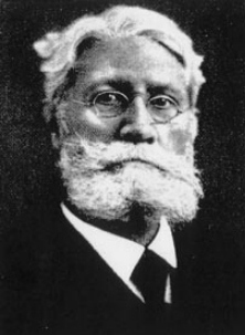 This image of Ludwig Courvoisier (1843-1918), who was a surgeon who described ‘Courvoisier’s law’ – ‘if in the presence of jaundice the gallbladder is palpable, then the jaundice is unlikely to be due to a stone.’ This was first published in his book ‘The pathology and surgery of the gallbladder’ in Leipzig in 1890. 54478 Origin of this photo from