Growth Time
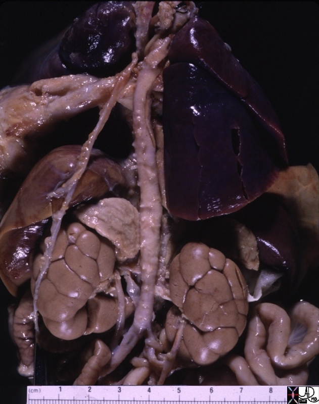
AGING OF THE PANCREAS In the young there is a tendency for the tail to be relatively large and the head to be relatively small. With age the reverse is true with the head appearing relatively large and the tail relatively small.
In general the pancreas loses acinar tissue as it ages and because it lacks a capsule, the fat of the surrounding tissues infiltrates and takes up the space of the atrophied acini. In general therefore the pancreas becomes more fatty with age.
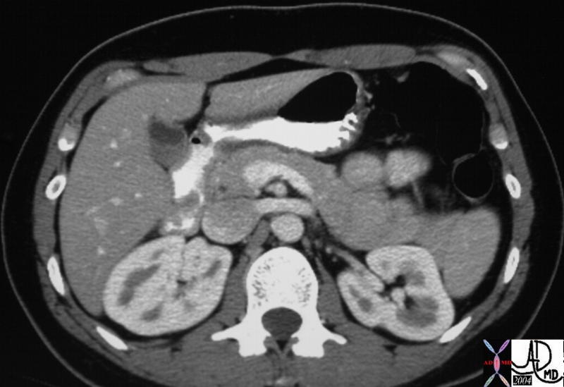
The CTscan through the body of the pancreas shows a calcification surrounded by a thin rim of soft tissue, just rightward of the poorly opacified SMV. The calcification represents a stone that is lodged in the distal CBD consistent with a diagnosis of choledocholithiasis. Incidental findings include the cysts in the right kidney. The pancreas has been almost totally replaced by fat which usually has no functional significance. Conclusion The pancreas as you have now learned is neither “all meat” nor a hermit. However it is an organ of duality with some unique structural features. As a gland it has both endocrine components as well as exocrine components. While the insulin secreting components only make up 2% of the gland, failure of their function leads to the devastating consequences and complications of diabetes mellitus. The pancreas has a dual blood supply and an extensive collateral arterial network ensuring that blood supply is always available, giving it the opportunity to keep its “finger on the pulse” of glucose metabolism. Diseases such as pancreatitis and carcinoma are not uncommon and the absence of a capsule results in early spread to surrounding organs with devastating results. Modern imaging has facilitated accurate evaluation of structural changes and so diagnosis and complications of disease can be accomplished with greater accuracy. The degree of spread of disease has significant impact on management and so better imaging has brought improved management. At this stage one can only wonder where imaging will take us next. We are in the mode now of combining functional imaging with structural imaging. We may not have a full sense of where the future of imaging will go but we do know that the imaging will always be based on structure, function, and pathology. It cannot be therefore overstated that it behooves us all to continue to acquire and retain a solid knowledge of anatomy, physiology and pathology. |

 This CT scan is from a relatively young male where the relative difference in size between the large tail and relatively small head is exemplified. Courtey Ashley Davidoff MD. a25138
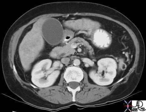 This CT scan through the pancreas in an older patient shows an atrophied body with a prominent head. 19387 CTscan Courtesy Ashley Davidoff MD
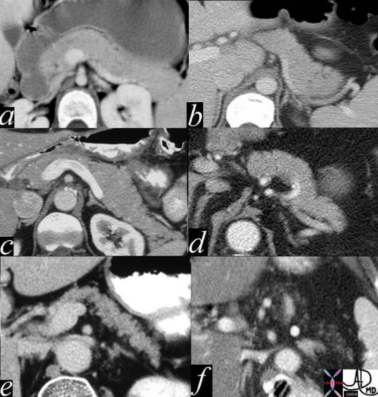 The size of the tail varies considerably between individuals, but in general it is relatively large in youth (a,b) starts to thin in middle age (c,d) and atrophies in the elderly (e,f) a= 12 year old female b= 30 year old female c= 30 year old male d= 40 year old male e= 70 year old female f = 80 year old female41394size02 Courtesy Ashley Davidoff MD
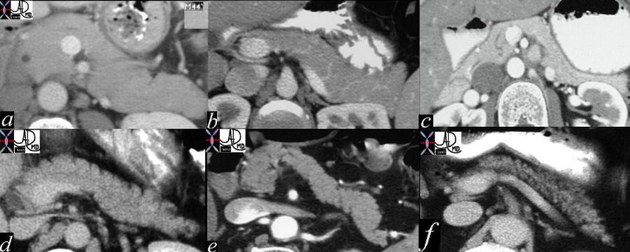 The variations of the size and surface shape of the body are noted in the above cases with the full and smooth body of youth in a, the gracile and smooth bodies of youth in b and c. Aging brings on atrophy and a wrinkling as evidenced by nodularity with a variety of manifestations. In d, the anterior border shows nodularity caused by early infiltration of fat , while in e atrophy is manifest with nodularity on both sides associated with thinning of the gland. In the last case (f) the nodularity and thinning caused by the fat infiltration is marked. 41505c02 Courtesy Ashley Davidoff MD
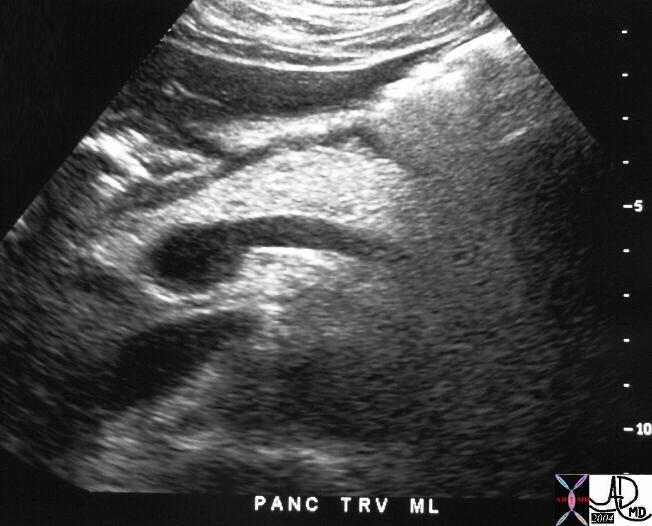 The transverse section through the pancreas shows an echogenic pancreas caused by infiltration of retroperitoneal fat into the age related involution of the pancreatic parenchyma. This is a normal and common finding,and usually has no clinical nor functional significance. 29495 Courtesy Ashley Davidoff MD